Eczema vs. Psoriasis: How to Tell Them Apart
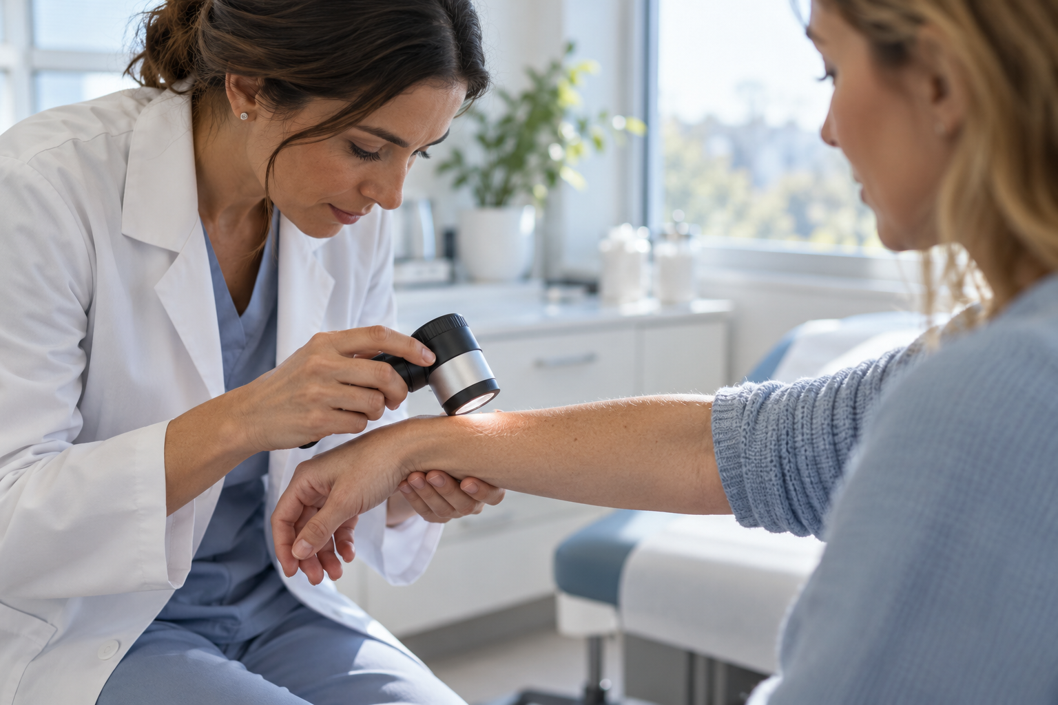
Patients walk into our office pointing at a patch of red, itchy skin and asking the same question: is this eczema or psoriasis? The confusion makes sense. Both conditions inflame the skin, both flare and fade over weeks or months, and both tend to show up in similar places on the body. Eczema and psoriasis come from different causes, though, and they respond to different treatments. Getting the right diagnosis changes what happens next, so our team at Aesthetic Dermatology Associates put together this guide to help you spot what separates one from the other.
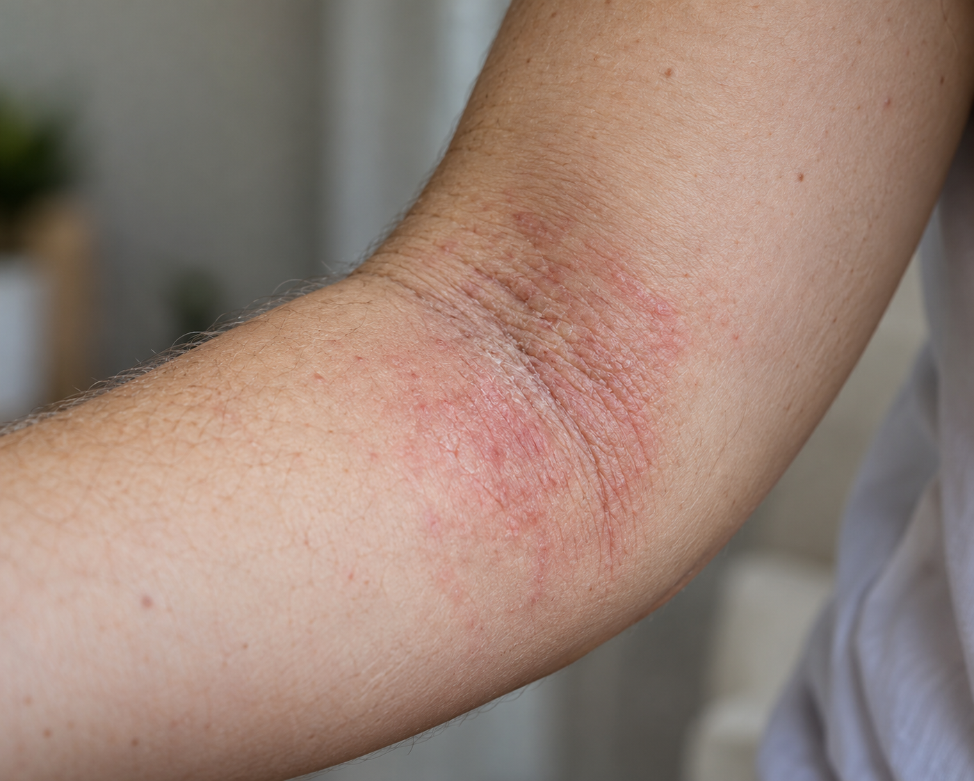
What Is Eczema?
Eczema, also known as atopic dermatitis, is a chronic condition rooted in how the skin barrier functions. There are many possible causes of eczema including genetic, environmental, skin barrier, and immune factors. People with eczema typically have a weakened skin barrier which can leave the skin open to irritants, allergens, and bacteria. The condition tends to run in families alongside asthma and seasonal allergies, and it commonly starts in childhood, though adults develop it too.
Weather changes, harsh soaps, stress, and rough fabrics can all trigger a flare. Once the skin barrier breaks down, inflammation follows, and the itch-scratch cycle takes over: scratching damages the skin further, which intensifies the itch, which leads to more scratching.
Eczema Symptoms
- Red or brownish-gray patches, depending on skin tone
- Intense itching, sometimes arriving before any visible rash
- Dry, cracked, or scaly texture
- Thickened skin from repeated scratching
- Small, fluid-filled bumps that ooze or crust when scratched
Common locations include the creases of the elbows and knees, the neck, wrists, however it can flare anywhere on the body. In infants, eczema often appears on the cheeks and scalp instead. The itch usually arrives first, before the skin visibly changes.
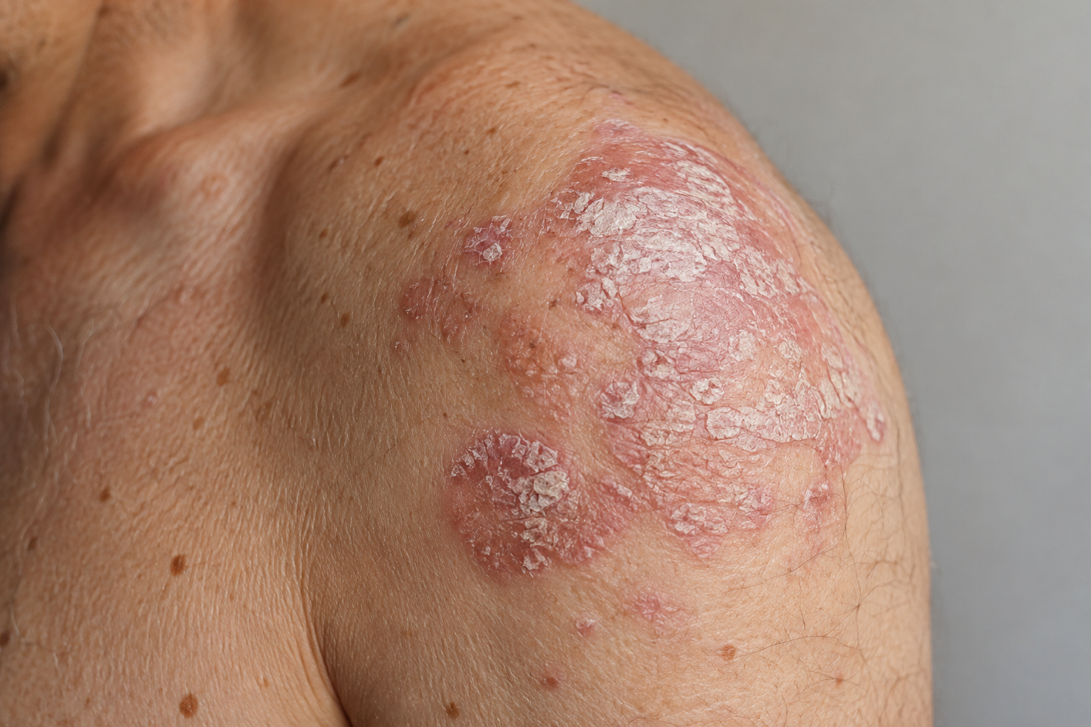
What Is Psoriasis?
Psoriasis works through a different mechanism. It’s an autoimmune condition: the immune system speeds up skin cell production, pushing cells through a cycle that normally takes about a month down to a matter of days. That buildup creates the thick, scaly patches psoriasis is known for.
Genetics play a large role, and psoriasis often first appears in early adulthood, though it can start at any age. Infections, skin injury, cold weather, and stress can all trigger a flare. Psoriasis can also affect the joints in some patients, a related condition called psoriatic arthritis, something eczema does not do.
Psoriasis Symptoms
- Raised patches with well-defined edges, called plaques
- A silvery-white scale layered over red or darkened skin
- Itching, burning, or soreness, depending on the patient
- Pitted, thickened, or discolored nails
- Patches that often mirror each other on both sides of the body
Common locations include the scalp, elbows, knees, and lower back. The scale and the sharp border around each plaque are two of the clearest visual differences from eczema.
Psoriasis vs. Eczema: The Key Differences
| Eczema | Psoriasis | |
|---|---|---|
| Cause | Multifactorial, including skin barrier dysfunction | Autoimmune, overactive cell turnover |
| Texture | Dry, cracked, sometimes oozing | Thick, raised, scaly plaques |
| Border | Poorly defined, blends into surrounding skin | Sharp, well-defined edges |
| Primary sensation | Intense itch | Itch, burning, or soreness |
| Typical locations | Elbow and knee creases, neck, wrists | Outer elbows, knees, scalp, lower back |
| Age of onset | Often begins in childhood | Often begins in early adulthood |
| Symmetry | Less consistent | Frequently symmetrical |
Texture usually settles the question. Eczema looks raw and irritated, while psoriasis builds up into a scale you can see sitting on the skin’s surface. Location matters too: eczema favors the folds where skin touches skin, psoriasis favors the outer surfaces of joints.
Skin doesn’t always follow the textbook, though. Some patients show features of both conditions, or a case that doesn’t fit either description cleanly. This is why we always recommend getting an in-person exam and evaluation.
Psoriasis Treatment
Psoriasis treatment depends on how much of the body it covers and how the plaques respond to earlier approaches. Our team considers a few paths:
- Topical treatments applied directly to plaques, used for mild to moderate cases
- Phototherapy, where controlled light exposure slows the overactive skin cell cycle
- Systemic treatments, used when psoriasis covers a large area or hasn’t responded to topical care and phototherapy
Eczema management is more focused on repairing the skin barrier, avoiding known triggers, and calming inflammation during flares. The two conditions come from different mechanisms, that’s why confirming the diagnosis comes before choosing a treatment plan.
When to See a Dermatologist
A rash that won’t clear with basic moisturizing, one that spreads, or one that disrupts sleep or daily activity is worth a professional look. Eczema and psoriasis can resemble other skin conditions too, including fungal infections and allergic reactions, so a photo or an internet search only goes so far. A dermatologist examines the pattern, texture, and history of the rash, and can confirm a diagnosis with a biopsy when the picture isn’t clear.
If you’re dealing with a persistent rash and aren’t sure what you’re looking at, our team at Aesthetic Dermatology Associates can help you get a clear answer and a plan that fits your skin.
Beyond the Breakout: Acne Scar Treatments That Work
By Catherine Waller, MD, board-certified dermatologist, Aesthetic Dermatology Associates

Acne likes to make its mark. While breakouts will eventually clear, the scars they leave behind often do not—at least not without help. Acne scarring can be a distressing, constant reminder of breakouts that are long gone. Fortunately, acne scars often can improve with the right treatment plan. When I discuss acne scars with my patients, I first and foremost ensure their acne is adequately controlled. Treating active acne is crucial to preventing further scarring.
Acne Scars Aren’t All the Same
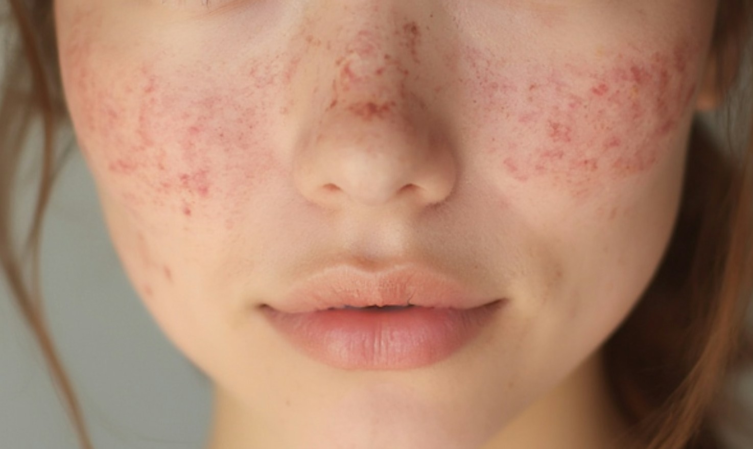
Before recommending a treatment, I look closely at the type of scarring present. Using the wrong treatment can mean spending time and money without getting the improvement you hoped for.
Icepick scars are narrow and deep, extending farther into the skin like a needle mark. Boxcar scars are wider depressions with well-defined edges and a flat or gently sloped base. Rolling scars create an uneven, wave-like appearance due to bands of scar tissue pulling the skin downwards. Hypertrophic and keloid scars sit raised above the skin and are the result of the body producing too much collagen during healing.
It is also important to differentiate true acne scarring from the reddish or brown discoloration that can follow a breakout. These marks are called post-inflammatory hyperpigmentation and do gradually fade with time and sun protection. True acne scars involve a structural change in the skin’s texture and often do not resolve without intervention.
Common Treatments for Acne Scarring
There is no one-size-fits-all approach. Most patients benefit from a combination of treatments rather than a single procedure. The best approach depends on the scar pattern, and different interventions address different aspects of scarring. The following procedures cover the scar types I see most often in practice, matched below to what each one treats:
Microneedling
Microneedling uses fine needles to create controlled micro-injuries in the skin. This stimulates the body’s wound-healing response and promotes new collagen formation, gradually improving the appearance and texture of depressed acne scars. Microneedling is particularly useful for many patients with rolling and boxcar scars and is considered safe for all skin tones. It is often performed as a series of treatments rather than a single session. Radiofrequency microneedling adds heat energy at the needle tip, which increases collagen remodeling and works well on deeper boxcar scarring. Like traditional microneedling, this can be used across a broad range of skin tones when performed with appropriate settings and technique.
Chemical peels
Peels use a controlled chemical solution to remove damaged outer layers of skin and prompt new cell turnover. Superficial and medium depth peels often can improve the appearance of superficial acne scars and post-inflammatory hyperpigmentation. Certain specialized chemical techniques can also be used for deeper, more focal icepick scars.
Laser resurfacing and light-based treatment
Laser treatment can address both scar texture and hyperpigmentation. Intense pulsed light targets discoloration without addressing texture, while resurfacing lasers work on the skin’s surface to soften the appearance of rolling and boxcar scars. These lasers carry a higher risk of dyspigmentation and therefore it is important that patients with melanin-rich skin see a provider who is trained in selecting the appropriate laser and settings for their skin tone.
Dermal fillers
Rolling and some boxcar scars sit low because the tissue underneath them has lost volume or become tethered to deeper layers. Filler placed directly under the scar lifts it to match the surrounding skin. The effect is temporary, lasting between six months and a year depending on the product, but it’s a fast option for patients who want visible improvement without a recovery period.
Intralesional Steroids
For hypertrophic and keloid scars we often consider injecting a steroid directly into the scar to help thin it out. This does carry a risk of hypopigmentation and thinning of the skin and should be done cautiously by a trained provider.
Surgery
Occasionally some hypertrophic or keloidal scars may also benefit from excision where the overgrown scar is replaced with a linear scar. This often will be paired with steroid injections to prevent the overgrown scar from reforming. Techniques such as punch excision can be used to replace some ice pick scars with a small linear scar. For rolling scars and certain tethered boxcar scars, subcision is a procedure designed to release bound-down scar tissue. Using a specialized needle or cannula beneath the skin, a dermatologist separates the scar tissue from the underlying structures that are tethering it down. Subcision is often combined with other treatments, such as microneedling, laser resurfacing, or dermal fillers, depending on the individual scar pattern.
What Determines Your Treatment Plan?
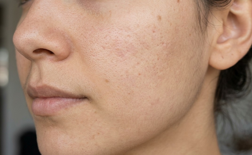
There is no single “best” acne scar treatment. The right approach depends on several factors. A patient’s skin tone determines which lasers and peels are safe to use. Scar type and depth also determine which modality or combination of treatments will work best. A plan built around one scar type won’t necessarily improve the other if both are present.
Downtime and budget matter too. A single dermal filler session takes about twenty minutes with no recovery time, while a series of resurfacing treatments generally requires planning around a week of visible healing between sessions. A treatment plan has to match the time and budget a patient can reasonably commit to.
What to Expect
Acne scar treatment is a process measured in sessions, not a single visit. Microneedling and radiofrequency microneedling typically require three to six sessions spaced a month apart to remodel collagen fully. Peels and light-based treatments often run in a series as well. Filler results are visible immediately but require maintenance appointments to sustain.
Improvement of 50 to 70 percent in scar depth and texture is a realistic goal for most patients who complete a full treatment course. Scar tissue is permanent structural change to the skin; treatment remodels and softens it rather than erasing it.
When to Book a Consultation
If your acne has been under control for a few months and what remains is texture, not active breakouts, that’s the time consider discussing acne scar treatments with a dermatologist.
At Aesthetic Dermatology Associates, we build treatment plans around your specific scar pattern and skin type rather than defaulting to one procedure for every patient. If you’re ready to talk through your options, book a consultation at our Media or Paoli office.
About the Author

Dr. Catherine Waller is a board-certified dermatologist at Aesthetic Dermatology Associates, treating the full spectrum of skin, hair, and nail conditions for patients of all ages. She earned her medical degree from Penn State College of Medicine and completed her residency at Penn State Milton S. Hershey Medical Center, where she served as chief resident. Her practice centers on personalized, evidence-based care, with a particular interest in skin cancer prevention and early detection. She is a member of the American Academy of Dermatology and the Pennsylvania Academy of Dermatology.
Related Links
Join Dr. Waller on LinkedIn professional network
See Dr. Waller’s provider page on our website
Dermal Fillers: Answering the Questions Our Patients Ask Most
By Leslie Sombers, Aesthetic Director, Aesthetic Dermatology Associates

I sit in on most filler consultations at our Paoli and Media offices. Patients want their cheeks fuller or their lines softer, but nearly every conversation starts with the same worry: “I don’t want to look like I had something done.” This article covers the questions patients ask most, pulled straight from those consultations.
What Are Dermal Fillers?
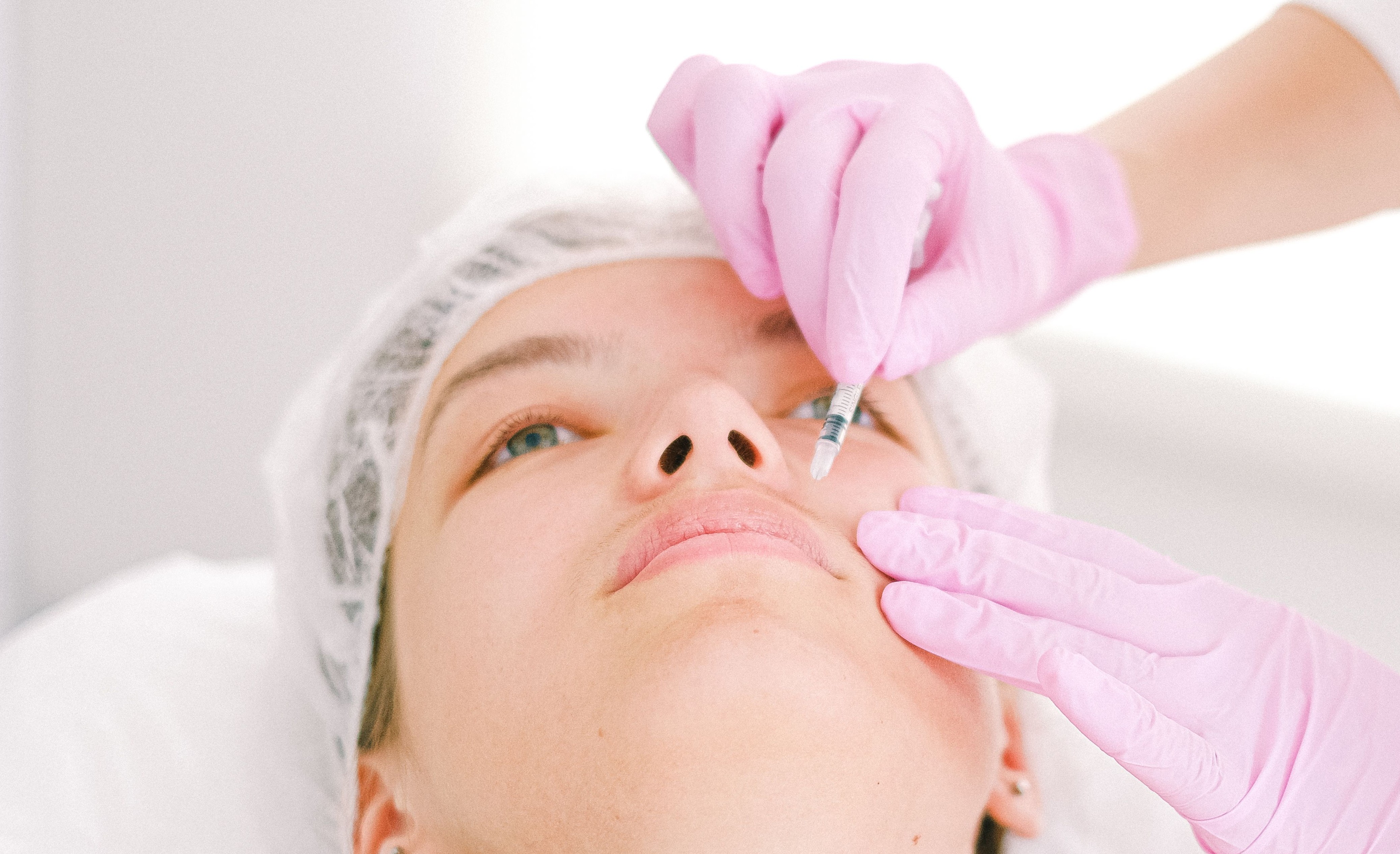
Dermal fillers are gel treatments, most made from hyaluronic acid, a substance your skin already produces to stay hydrated and full. A physician injects the filler beneath the skin to restore lost volume, soften lines, or add definition to the lips or cheeks. Sessions take under an hour. No surgery, no incisions, and most patients return to normal activities the same day.
The Dermal Filler Procedure: What to Expect
Your physician starts with a facial assessment. They study your bone structure, skin quality, and the areas that concern you, then build a treatment plan designed to support your unique facial structure.
Before injecting, we clean the treatment area and apply numbing cream. Most fillers we use also contain lidocaine, which reduces discomfort during injection. Your physician places the filler in small amounts, carefully assessing symmetry and balance throughout the treatment to create a natural result.
Most patients describe the sensation as pressure more than pain. Expect mild swelling or bruising at the injection sites for a few days. You will see initial results of the treatment the same day, and as the swelling settles after a week or two, you can see the final result.

Will It Look Natural, or Will I Look Overdone?
This is the question we hear most, and it shapes how our physicians practice.
“Our physicians treat conservatively. When I do a consult, it is more about facial balancing, a much more natural and conservative approach.”
Facial balancing means looking at your whole face, not one feature in isolation. Your physician might add volume to a hollow cheek to soften a nasolabial fold without touching it directly, or restore definition to your lip border instead of overfilling the center of the lip. The goal at every consult: a result your friends notice as “you look great” or “you look rested.”
Dermal Fillers: Pros and Cons
Filler offers real benefits, and it comes with trade-offs worth knowing before you book a consult.
Pros:
- Results show up the same day, with no recovery time built into your schedule
- Hyaluronidase can dissolve hyaluronic acid filler, so the treatment is reversible
- Sessions run 30 to 45 minutes in most cases
- Placed conservatively, filler restores volume that compliments your overall facial structure
Cons:
- Results fade over time and require maintenance appointments
- Bruising and swelling can occur and take several days to resolve
- There are costs associated with each treatment, and it may be different each time depending on what is recommended
- Poor technique or the wrong product for an area can produce undesired results, which is why physician experience matters as much as the product
How Long Do Results Last?
Duration depends on the area treated, the product used, and how fast your body breaks down hyaluronic acid. Areas with more movement, like the lips, tend to metabolize filler faster than areas like the cheeks. Most patients see results last several months to a year before they need a touch-up. Your physician will give you a specific estimate based on your treatment plan at your consult.
Where Does Filler Go, and Does It Ever Fully Dissolve?
Hyaluronic acid filler doesn’t sit under your skin waiting to be removed. Your body breaks it down over time through the same metabolic process it uses for the hyaluronic acid your skin makes on its own. Results fade over time instead of disappearing all at once, which is why most patients return for a touch-up.
You may have seen claims online that filler never fully dissolves and migrates to other parts of the face, creating unnatural results months or years later. True migration exists, but it’s uncommon. Overfilling an area, using the wrong product for that location, or layering repeated treatments without accounting for what’s already there account for most cases. Technique and injected volume, more than the filler itself, drive that outcome.
Because of this, our physicians map out a plan based on your anatomy before picking up a syringe. Small, precise amounts placed in the right structures stay where they’re placed and break down predictably.
I’ve Lost Weight: Can Filler Help with Volume Loss (Ozempic Face)?
Rapid weight loss, including from GLP-1 medications like Ozempic and Wegovy, tends to show up in the face before anywhere else. Fat loss in the cheeks and temples can leave skin looking hollow or aged, even when the rest of the body looks toned and fit.
Filler restores volume in the cheeks, temples, and other areas that have lost fat. Many patients who’ve gone through significant weight loss find that a conservative amount of filler brings back a rested appearance without adding weight anywhere else. If you’ve lost weight and don’t recognize your own face in photos, that’s worth raising at a consult.
How Much Does It Cost?
Filler pricing depends on the areas treated, the amount of product used, and your specific goals, so we don’t quote prices without seeing you first. At your consult, your physician maps out a treatment plan and gives you an exact cost before you decide anything. You’re under no obligation to move forward after that conversation.
Where We Treat (and Where We Don’t)
We treat the nasolabial folds, marionette lines, cheeks, tear troughs, and lips more than any other areas. These are the spots where volume loss shows up early and where filler makes a visible, natural difference.
We treat the chin, jawline, and temples far less. Filler can work in those areas, but our consults start with what your face needs, not a checklist of every place filler could theoretically go. If your consult points to one of those areas, we’ll discuss it. If it doesn’t, we won’t push it.
Dermal Filler Aftercare: Tips for a Smooth Recovery
Aftercare is straightforward, and following it cuts down on bruising, swelling, and downtime.
- Skip strenuous exercise for 24 hours after treatment. Increased blood flow can worsen swelling and bruising.
- Avoid alcohol for 24 hours before and after your appointment. Alcohol thins your blood and raises your bruising risk.
- Ice the treated area in short intervals during the first day if you notice swelling.
- Sleep on your back for the first night when you can, especially after lip or cheek treatment.
- Hold off on facials, massages, or other pressure on the treated area for about two weeks.
- Skip saunas, hot yoga, and heavy sun exposure for a few days after treatment.
- Call our office if you notice unusual pain, skin that turns pale or white, or symptoms beyond typical swelling and bruising. These are rare, but we want to know right away.
A Final Word from Leslie

Every patient who sits down with one of our physicians wants the same thing: a well-rested version of themselves, not a different person. We hold ourselves to that standard at every consult, and we’d rather treat conservatively and adjust over time than overcorrect in one visit.
If you have questions about dermal fillers that we haven’t covered here, bring them to your consult. That conversation, not this article, is where your treatment plan takes shape.
Punch Biopsy vs Shave Biopsy: Healing Stages, Scarring, and What to Expect from Each
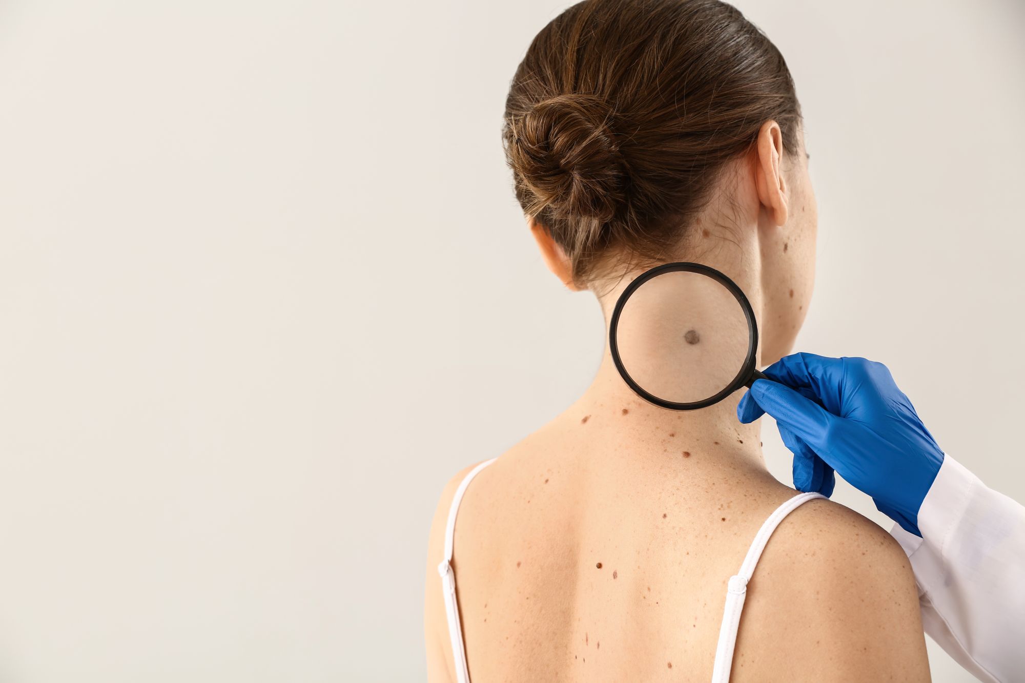
When your dermatologist recommends a skin biopsy, the conversation often centers on what the results might reveal — not on what the healing process will actually look like. But the type of biopsy you have matters, and it matters quite a bit when it comes to how your skin heals, how long recovery takes, and whether you’re likely to have a scar.
The two most common biopsy types performed at Aesthetic Dermatology Associates are the shave biopsy and the punch biopsy. Though both remove a small sample of skin for laboratory analysis, the technique, depth, and wound each type leaves behind are meaningfully different — which is why the healing stages and aftercare instructions are different too.
This article walks you through each type so you know exactly what to expect from the moment you leave our Paoli or Media office through the weeks that follow.
Shave biopsy: what it is and when it’s used
A shave biopsy uses a thin, flexible blade to remove a raised or surface-level lesion — shaving it off at or just below skin level, typically without the need for sutures. It is the most commonly used technique for evaluating moles, skin tags, seborrheic keratoses, and surface-level lesions suspected of being basal cell carcinoma or squamous cell carcinoma.
Because the blade does not go deep into the dermis, the wound is shallow and saucer-shaped — think of a small, clean scrape rather than a puncture. This characteristic determines everything about how it heals.
Punch biopsy: what it is and when it’s used
A punch biopsy uses a small circular blade — ranging from 2mm to 6mm in diameter — to remove a cylindrical core of skin that extends through all layers of the dermis and into the subcutaneous tissue below. The resulting wound is round and deeper than a shave biopsy, and typically requires one or two sutures to close.
Punch biopsies are used when your dermatologist needs a full-thickness tissue sample to evaluate inflammatory skin conditions such as eczema or psoriasis, deeper pigmented lesions, suspected melanoma, or cases where a more complete tissue architecture is needed for an accurate diagnosis.
Shave biopsy: healing stages
Understanding the shave biopsy healing process ahead of time helps remove the worry from what can look alarming in the first week.
Days 1–3: the wound is raw and may crust
The site will look red, shallow, and raw — similar in appearance to a minor abrasion. A thin crust or scab may begin to form within the first day or two. This is normal. Keep the area moist with a thin layer of petrolatum (Vaseline or Aquaphor) and covered with a non-stick bandage, changing it once daily. Do not let the wound dry out — moisture supports faster, cleaner healing and reduces scarring.
Days 4–10: new skin begins to form
The crust gradually lifts at the edges as new skin forms underneath. The area may be slightly pink or lighter than the surrounding skin. Resist the urge to pick at any scab — removing it prematurely disrupts the healing layer beneath and increases the likelihood of a more visible scar.
Weeks 2–4: the surface closes
By the end of the second week, most shave biopsy sites have re-surfaced. The area will appear pink or mildly discolored — often lighter or slightly darker than your surrounding skin — but the wound itself is closed. You can usually reduce bandaging at this stage, though you should continue sun protection over the area. Ultraviolet exposure on a healing biopsy site significantly increases the risk of persistent discoloration.
Months 2–6: color and texture normalize
Shave biopsy sites often leave a subtle, flat, lighter or pink mark that fades significantly over several months. On areas with a good blood supply — the face, for instance — healing and fading tend to be faster. On the trunk or legs, color normalization can take longer. Diligent sun protection during this window makes a meaningful difference in the final result.
Punch biopsy: healing stages
Because a punch biopsy goes deeper and is typically sutured, the healing stages look and feel different from a shave biopsy.
Days 1–3: sutured wound, some bruising and swelling
The closed wound will feel slightly raised and tender. Light bruising and swelling around the sutures is normal. Keep the area clean, dry, and protected. Your dermatologist will give you specific instructions, but the general approach is the same: petrolatum, a non-stick dressing, and once-daily changes.
Days 7–14: suture removal
Sutures are typically removed between 7 and 14 days depending on the location — earlier on the face (where healing is faster) and later on the back or legs (where healing is slower and tension is higher). After suture removal, the linear or oval scar will be pink and slightly raised. This is the early scar-formation phase and does not reflect the final result.
Weeks 3–8: scar softening
Over the following weeks, the punch biopsy scar flattens and begins to soften. It may remain red or pink during this period — this is normal vascularization as the tissue remodels. Keeping the area moisturized and out of direct sun significantly helps the scar mature more smoothly.
Months 3–12: final scar appearance
Punch biopsy scars generally mature into a small, flat, round or oval mark. On the face, well-closed punch biopsies often become barely visible over time, particularly when the wound edges were aligned carefully at suturing. On the trunk and extremities, the scar tends to remain more visible but continues to fade for up to a year. Scar creams containing silicone can be used after the wound is fully closed to support the remodeling process.
Scarring: shave vs punch biopsy
This is the question most patients ask, and the honest answer is that both types of biopsies can leave a mark — but the character of that mark is different.
Shave biopsies typically leave a flat, smooth, slightly lighter or mildly discolored area that blends into surrounding skin over months. Punch biopsy scars are more defined in shape — a small round or oval mark — and go through a more visible maturation process. In experienced hands, both can have excellent cosmetic outcomes, particularly on the face.
Location matters enormously. Biopsy scars on the nose, cheek, or forehead tend to heal with better cosmetic results than those on the chest, shoulders, or upper back, where wound tension is higher and the skin heals differently. If you had a skin cancer biopsy on your face or nose, be patient — the site will look worse before it looks better, and the final result at six months is almost always more reassuring than it appears at three weeks.
Mole biopsy healing: which type applies to you
When a mole is biopsied, either technique may be used depending on its characteristics. Raised, dome-shaped moles are typically sampled with a shave biopsy. Flat, atypical, or deeply pigmented moles — particularly those being evaluated for melanoma — are more likely to require a punch biopsy to obtain a full-thickness sample.
If you’re uncertain which type of biopsy you had, look at the wound: if it was sutured, it was a punch biopsy. If there were no sutures and the wound looks like a scrape, it was a shave biopsy.
Mole biopsy healing follows the same general stages described above for each type. The main difference is that because moles are often raised, the shave biopsy site may look slightly sunken after the lesion is removed — this typically fills in over the first few months.
Skin cancer biopsy: does the type affect what comes next?
If your biopsy was performed to evaluate a lesion for skin cancer — whether basal cell carcinoma, squamous cell carcinoma, or melanoma — the type of biopsy used is chosen to give your dermatologist the diagnostic information they need, not primarily for cosmetic reasons. A basal cell biopsy is often a shave sample, since BCCs tend to be surface-level. Melanoma evaluation frequently requires a punch or excisional biopsy to assess depth.
A positive skin cancer biopsy result means a further conversation about treatment — which may include excision, Mohs surgery, or superficial radiation therapy depending on the type, location, and depth. The biopsy site itself may not be the final treatment site; your dermatologist will discuss margins and next steps at your follow-up appointment.
If you are waiting for skin cancer biopsy results, standard turnaround from the dermatopathology lab is typically 5 to 14 business days. Results for basal cell and squamous cell carcinoma often return closer to the shorter end of that range.
Signs a biopsy site is not healing normally
Most biopsy sites heal without complications. However, contact Aesthetic Dermatology Associates if you notice any of the following:
- Increasing redness, warmth, or swelling beyond the first two to three days
- Pus or discharge with an odor
- Fever or red streaks extending from the wound site
- A wound that appears to be enlarging rather than closing
- Sutures that come loose before your scheduled removal appointment
- A site that is not healing after four to six weeks and remains open or raw
Infection after a skin biopsy is uncommon but treatable. Don’t wait to contact us — catching it early means a simpler course of treatment.
Frequently asked questions
How long does a shave biopsy take to heal?
Most shave biopsy sites are fully re-surfaced within 2 to 3 weeks. Color normalization takes longer — typically 2 to 6 months depending on your skin tone and the location of the biopsy.
How long does a punch biopsy take to heal?
The wound closes by 2 weeks (after suture removal). The scar continues to mature for 3 to 12 months. Most patients see a significantly improved appearance by 3 months.
Will my punch biopsy scar be noticeable?
Punch biopsy scars are typically small and round, and fade considerably over time. On the face, they are often nearly imperceptible at one year. On the trunk or extremities, they tend to remain more visible but continue to soften. Sun protection during healing is the single most important thing you can do to minimize the final appearance.
How long should I keep the biopsy site covered?
For shave biopsies, continue covering the site with a petrolatum-coated bandage until the wound is fully re-surfaced — typically 10 to 14 days. For punch biopsies, keep the site covered and protected until suture removal, then follow your dermatologist’s guidance for the weeks after.
Can I exercise after a skin biopsy?
Light activity is generally fine within a day or two. Avoid vigorous exercise, heavy sweating, or activities that put direct tension on the biopsy site for at least the first week, particularly for punch biopsies — elevated heart rate and sweating can cause bleeding, and movement tension on sutured wounds can disrupt healing.
Have questions about your biopsy or results?
If you’ve had a skin biopsy at Aesthetic Dermatology Associates and have questions about how your healing is progressing, or if you’re waiting for results and would like to speak with a member of our clinical team, our Paoli and Media offices are here to help. You can reach us by calling either location directly.
And if you haven’t yet had a skin check but have a spot or mole you’re concerned about, now is a good time to schedule one. Most skin cancers are highly treatable when caught early — a brief appointment with one of our dermatologists is the simplest way to get an answer.
Medical Dermatology in Paoli, PA: What to Expect at a Board-Certified Dermatology Practice
If you’ve been searching for a dermatologist in Paoli, PA — or looking for dermatology care serving Malvern, Wayne, Berwyn, Exton, or West Chester — you may already know that not all dermatology practices offer the same scope of care. Some focus primarily on cosmetics. Others handle only routine skin checks. A full-service medical dermatology practice does something more: it treats the conditions that affect your skin’s health, function, and long-term wellbeing.
At Aesthetic Dermatology Associates in Paoli, our board-certified dermatologists and clinical team provide comprehensive medical dermatology care for patients throughout Chester County and the Main Line. This article walks through what that looks like in practice — what conditions we treat, how we approach them, and what to expect as a patient.
What Is Medical Dermatology?
Medical dermatology refers to the diagnosis and treatment of skin conditions that have a clinical, not purely cosmetic, basis. This includes everything from chronic conditions like eczema and psoriasis to acute concerns like acne, to serious diagnoses like skin cancer.
While cosmetic dermatology addresses how the skin looks, medical dermatology addresses how the skin functions — its barrier integrity, its immune response, its susceptibility to disease. The two often overlap: a patient being treated for skin cancer may also have cosmetic concerns about scarring; a patient managing psoriasis may struggle with self-confidence. Good dermatological care acknowledges the whole picture.
Conditions We Treat at Our Paoli Office
Skin Cancer
Skin cancer is the most common type of cancer in the United States — and one of the most treatable, when caught early. At Aesthetic Dermatology Associates, skin cancer care begins with thorough full-body skin exams to identify suspicious lesions, followed by biopsy when indicated and a treatment plan tailored to the type and stage of cancer.
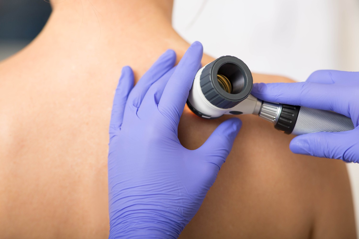
The most common types we diagnose and treat are basal cell carcinoma (BCC) and squamous cell carcinoma (SCC). Melanoma, while less common, is the most serious form and requires prompt attention when detected.
For patients throughout the Paoli and Main Line area, early detection is everything. If you haven’t had a full-body skin exam recently — particularly if you’ve had significant sun exposure over your lifetime, a family history of skin cancer, or a personal history of sunburns — scheduling a screening is one of the most impactful things you can do for your long-term health.
Photodynamic Therapy (PDT)
Photodynamic Therapy is a light-activated treatment that targets sun-damaged and precancerous skin while protecting surrounding healthy tissue. It’s one of the most effective non-surgical options for treating actinic keratoses — the rough, scaly patches that develop after years of UV exposure and can progress to squamous cell carcinoma if left untreated.

Image: PDT Device used in our clinics
The treatment involves applying a photosensitizing agent to the skin, allowing it to absorb into abnormal cells, then activating it with a specific wavelength of light. The result is targeted destruction of damaged cells with minimal impact on healthy surrounding tissue.
PDT is particularly well-suited for patients with widespread sun damage across the face, scalp, or forearms — areas where treating each lesion individually would be impractical. It also offers a cosmetic benefit: many patients notice an improvement in overall skin tone and texture following treatment.
For patients coming from Malvern, Berwyn, Wayne, and surrounding areas, PDT is available at our Paoli office without the need for hospital visits or surgical settings.
Dermatologic Surgery
When a skin lesion needs to be removed — whether a concerning mole, a cyst, a skin cancer, or a growth causing discomfort — dermatologic surgery provides a precise, effective solution. All surgical procedures at our Paoli practice are performed comfortably in our office with local anesthesia, so you can return home the same day.
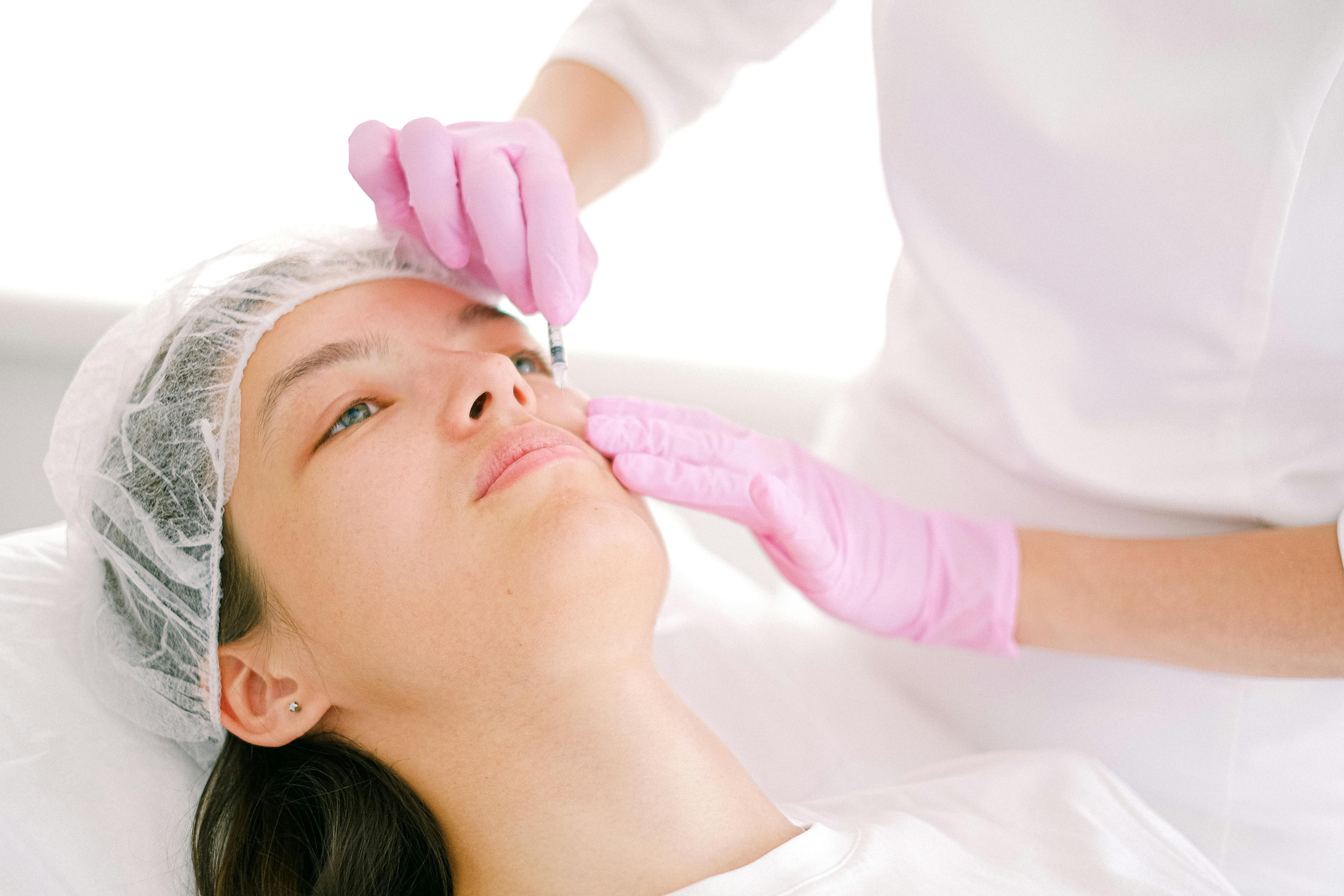
There’s no need for a hospital, a surgical center, or general anesthesia. Most patients describe the procedure itself as far less intimidating than they expected — the local anesthetic does its work, and the process is straightforward.
Common dermatologic surgeries we perform include excision of skin cancers, removal of cysts and lipomas, and treatment of other growths that require histopathological evaluation. Each specimen removed is sent for pathology analysis to confirm complete removal and accurate diagnosis.
For patients in Exton, West Chester, and across Chester County who’ve been referred for a procedure, our office offers a calm, clinical environment close to home.
Acne Treatment
Acne is one of the most common skin conditions we treat — and one of the most misunderstood. It’s not a teenage problem that resolves on its own, and it’s not simply a matter of better skincare. Acne is a medical condition with multiple contributing factors: excess sebum production, bacterial overgrowth, inflammation, hormonal influence, and genetics all play a role.

At Aesthetic Dermatology Associates, we provide evidence-based treatments to manage and reduce acne effectively — not just manage outbreaks as they occur, but address the underlying drivers. That may include topical or oral prescription medications, hormonal therapy where appropriate, in-office procedures, or a combination.
We also take scarring seriously. Acne that’s inadequately treated — or that’s been present for years — can leave lasting marks. Addressing active acne early, aggressively, and correctly is the best way to protect the skin’s long-term appearance and health.
Patients searching for a dermatologist in Paoli or Malvern for acne care will find that we approach this condition with the same clinical seriousness we bring to any other diagnosis.
Eczema
Eczema — or atopic dermatitis — is a chronic, non-contagious condition that causes dry, itchy, and inflamed skin. It’s particularly common in children, but it affects patients of all ages, and many adults live with eczema for decades without receiving adequate treatment.
The condition is driven by a compromised skin barrier that allows moisture to escape and irritants to penetrate. This triggers an immune response that causes the characteristic redness, itching, and flaking. In moderate to severe cases, it can significantly affect sleep, concentration, and quality of life.
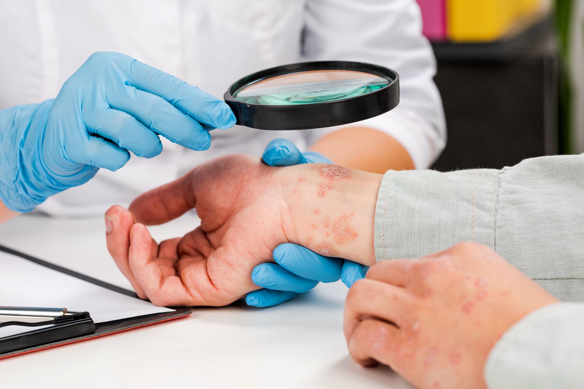
Effective eczema management involves more than a hydrocortisone cream. Our approach addresses the skin barrier directly — with appropriate moisturization protocols, trigger identification, and prescription treatments that range from topical corticosteroids and calcineurin inhibitors to newer targeted biologics for patients with moderate to severe disease.
For families in Wayne, Berwyn, and surrounding communities looking for a dermatologist who will take a chronic eczema diagnosis seriously rather than issue a repeat prescription, our Paoli office offers the continuity of care this condition requires.
Psoriasis
Psoriasis is a chronic autoimmune skin condition characterized by itchy, scaly patches that most commonly appear on the torso, elbows, knees, and scalp. Beyond the visible symptoms, psoriasis can impact sleep, focus, and overall comfort — and is associated with systemic inflammation that carries implications beyond the skin, including increased cardiovascular risk and a condition called psoriatic arthritis.
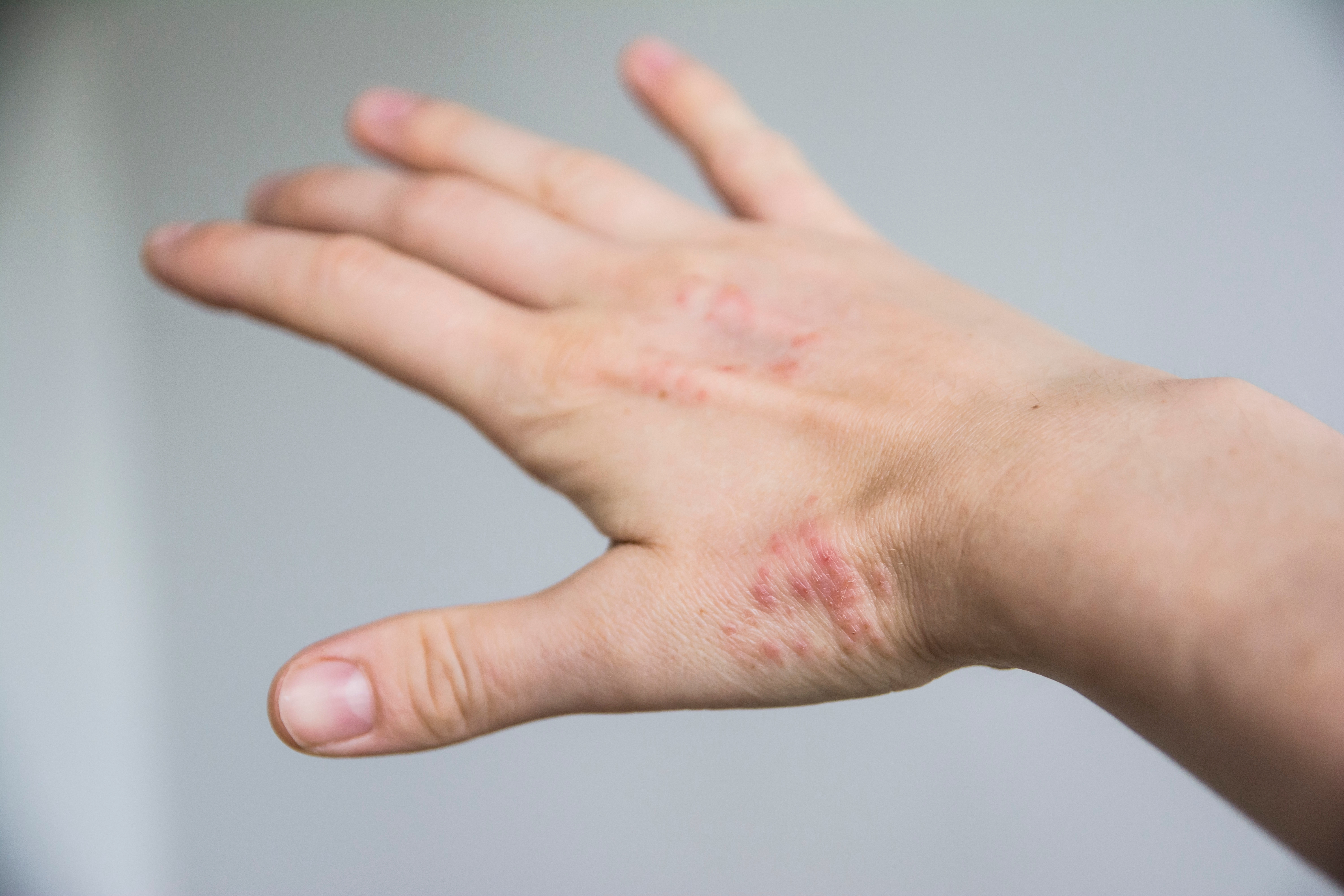
This is a condition that benefits significantly from specialist management. General practitioners can offer basic treatment, but a board-certified dermatologist brings a broader range of treatment options — including topical therapies, phototherapy, oral systemic medications, and biologics that have transformed outcomes for moderate to severe psoriasis patients over the past decade.
If you’ve been managing psoriasis on your own, or if previous treatment hasn’t given you adequate control, a consultation at our Paoli office is a practical next step. We serve psoriasis patients from across the Main Line, including Malvern, Exton, and West Chester.
Mole Removal and Evaluation
Moles are pigmented cell clusters that vary in size, shape, and color — most are entirely harmless and require no intervention. But changes in a mole’s appearance deserve medical attention. The classic warning signs follow the ABCDE framework: Asymmetry, Border irregularity, Color variation, Diameter greater than 6mm, and Evolution over time.
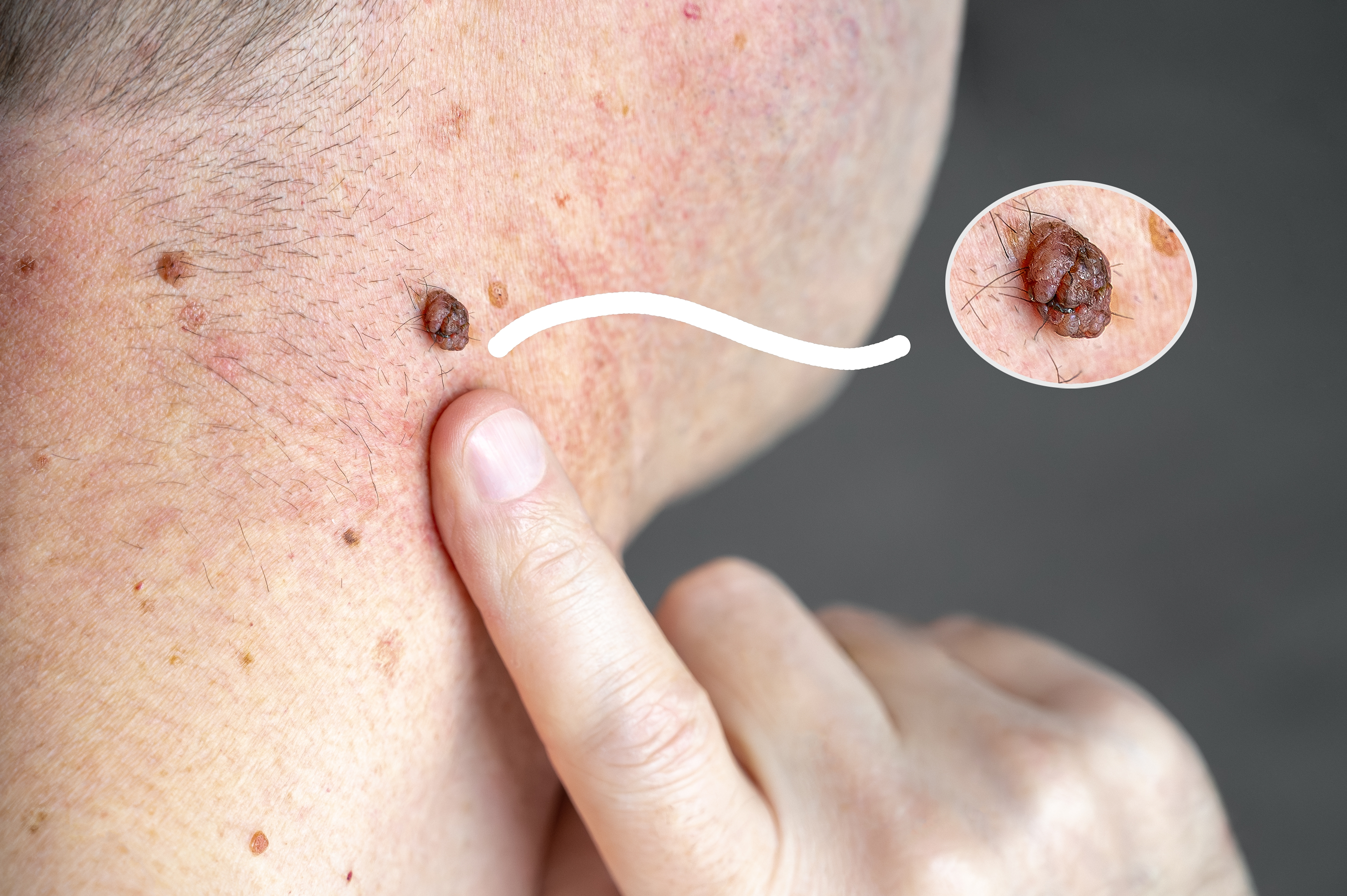
If you’ve noticed a mole changing, itching, bleeding, or simply looking different than it did before, the appropriate first step is a clinical evaluation — not a wait-and-see approach.
At Aesthetic Dermatology Associates, mole evaluation is part of routine full-body skin exams. When a mole warrants removal — either for diagnostic purposes or because it’s causing concern — we perform the procedure in-office with minimal discomfort and send all specimens to pathology.
Why See a Dermatologist?
Many skin conditions are best managed by a board-certified dermatologist. Dermatologists complete four years of residency training specifically focused on the skin, hair, nails, and mucous membranes, plus any additional fellowship training. They have the clinical experience to identify conditions that may look similar on the surface but require very different treatment approaches.
For patients in Paoli, Malvern, Wayne, Berwyn, Exton, and West Chester, having a board-certified dermatologist accessible on the Main Line — rather than requiring a trip into Philadelphia — makes regular skin care practical rather than something that gets postponed indefinitely.
What to Expect at Your First Appointment
New patients at Aesthetic Dermatology Associates are seen for a thorough consultation. Depending on your concerns, this may include a full-body skin exam, evaluation of specific lesions or conditions you’ve noticed, a review of your skin and family history, and a discussion of treatment options.
We don’t rush appointments. Medical dermatology requires clinical judgment, and clinical judgment requires time — to look carefully, to ask the right questions, and to explain findings in a way that makes sense to you.
If you’re coming to us from Paoli, Malvern, Wayne, Berwyn, Exton, West Chester, or anywhere across Chester County and the Main Line, you’re welcome to request an appointment online or call our office directly.
How to Perform a Skin Cancer Self-Exam: What Dermatologists Look For
Featuring Dr. Clare Goggins, a board-certified dermatologist providing expert medical care to both adult and pediatric patients.
You’ve noticed something on your skin that wasn’t there before. Or maybe there’s a mole that looks different than it used to. So you’re doing what most people do at 11 PM on a Tuesday: searching the internet, comparing what you’re seeing to images online, and quietly spiraling into worst-case scenarios.
If you’re here because you’re worried about skin cancer, let’s start with this: noticing changes in your skin and taking them seriously is exactly what you should be doing. Skin awareness isn’t paranoia—it’s smart healthcare.
Dr. Clare Goggins, a board-certified dermatologist at Aesthetic Dermatology Associates, sees patients every week who are anxious about skin changes they’ve discovered. Her approach combines Johns Hopkins-trained clinical expertise with something equally important: helping patients understand what they are actually looking at.
“The goal of self-examination isn’t to diagnose yourself,” Dr. Goggins explains. “It’s to become familiar enough with your own skin that you notice when something changes. That awareness—combined with regular professional screenings—is what catches skin cancer early, when it’s most treatable.”
Why Self-Exams Matter (But Can’t Replace Your Dermatologist)
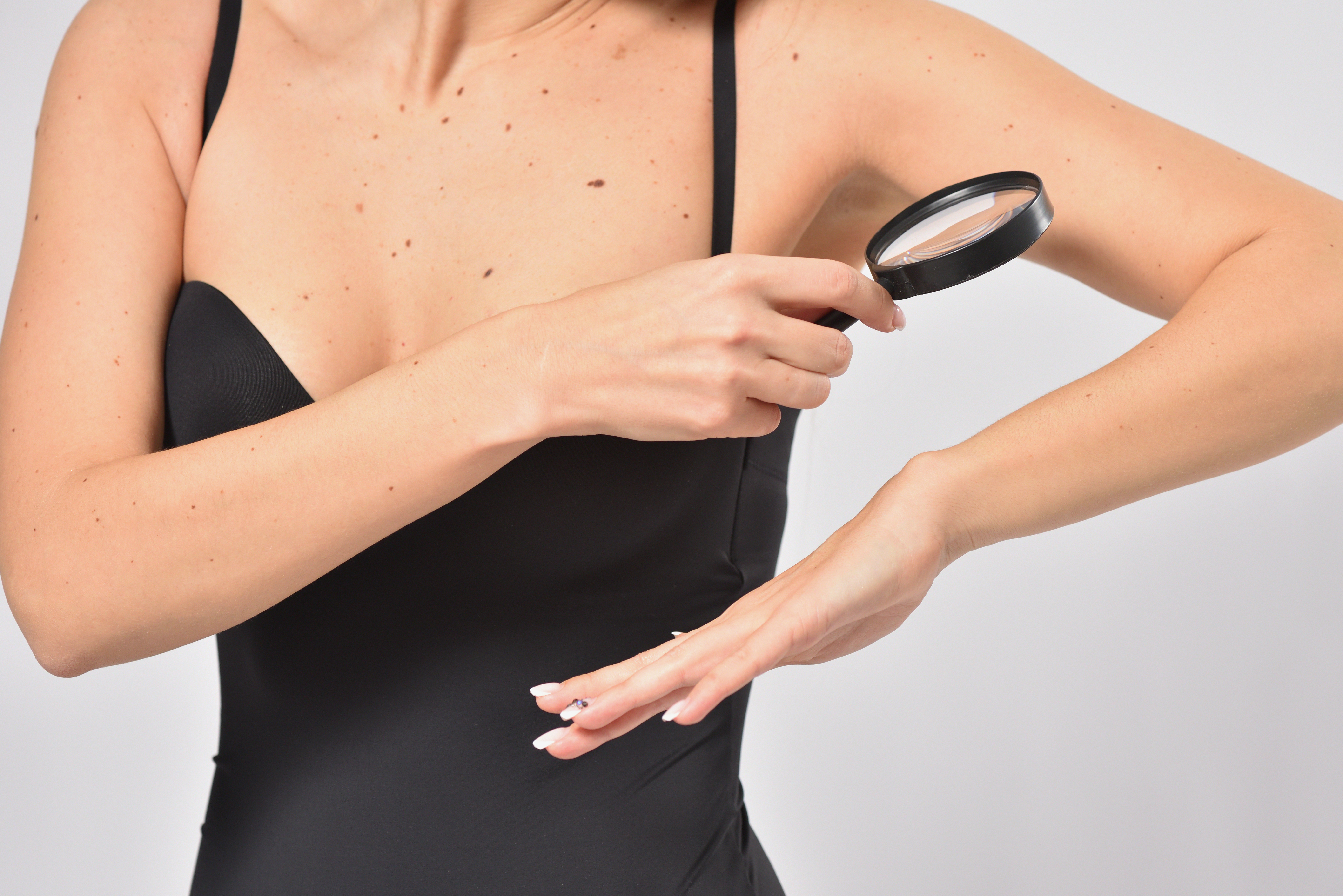
Here’s something that might surprise you: most skin cancers are first noticed by patients themselves, not by doctors during routine exams. You see your skin every single day. Your dermatologist sees it once, maybe twice, a year depending on your medical history.
But here’s the critical distinction: self-exams help you know when to see a doctor. They don’t tell you what something is or isn’t. That anxiety you’re feeling while comparing your mole to images online? That’s your cue to stop searching and schedule an appointment instead.
The ABCDE Method: What Dermatologists Look For
Dermatologists recommend a framework called the ABCDE method to identify suspicious moles and melanoma. This is the same clinical approach Dr. Goggins uses when examining patients.
Asymmetry: If you drew a line through the middle of the mole, would both halves look the same? Normal moles are typically symmetrical. Melanomas often aren’t.
Border irregularity: Normal moles have smooth, even edges. If the border is ragged, notched, blurred, or irregular, that’s worth noting.
Color variation: A normal mole is usually one uniform color. If you’re seeing multiple colors within the same spot (brown mixed with black, red, white, or blue), that’s a warning sign.
Diameter: Melanomas are often (though not always) larger than 6mm—about the size of a pencil eraser. But don’t discount something just because it’s small.
Evolving: This is the most important letter. Has the mole changed over the past few weeks or months? Is it getting bigger, darker, raised, or behaving differently? Change over time is the single biggest red flag.
Dr. Goggins adds an important caveat: “The ABCDE method is incredibly useful, but it’s not foolproof. That’s why the ‘ugly duckling’ sign matters too—if you have one mole that just looks different from all your other moles, even if it doesn’t check the ABCDE boxes, call your dermatologist to have it examined.”
How to Perform a Self-Exam (Step by Step)
You don’t need special equipment or medical training. You need decent lighting, two mirrors (a full-length mirror and a handheld one), and about 15 minutes of uninterrupted time. Do this once a month.
Start at your face and scalp. Examine your face, ears, neck, and scalp carefully. Use the handheld mirror to check areas you can’t see directly. If you have thick hair, use a blow dryer to part sections so you can see your scalp.
Move down your body systematically. Check your shoulders, chest, and torso—front and back. Look under your arms. Examine both sides of your arms, your hands, between your fingers, and under your nails.
Check areas that don’t see the sun. Look at your buttocks, genital area, and the backs of your legs using your mirrors. Melanoma doesn’t only develop on sun-exposed skin—it can appear anywhere.
Don’t skip your feet. Check the tops, bottoms, between your toes, and your toenails.
Optional but useful: Take photos. If you have a lot of moles or spots you’re monitoring, taking monthly photos can help you track changes over time more objectively than memory alone.
What To Look For: Warning Signs vs. Normal Findings
Things that usually warrant a call to your dermatologist:
- A new growth appearing after age 30, especially one that’s growing or changing
- Any mole that looks noticeably different from your other moles (the ugly duckling sign)
- A sore that doesn’t heal within 3-4 weeks
- A spot that repeatedly itches, bleeds, crusts over, or oozes
- A mole that’s changing in size, shape, color, or texture
- A dark streak appearing on a fingernail or toenail
Common benign findings that may catch your attention:
Seborrheic keratoses look like brown or black growths stuck onto the skin’s surface. They’re extremely common as you age and can look concerning because they’re often dark and crusty, but they’re harmless.
Cherry angiomas are small, bright red bumps that appear with age. They’re clusters of blood vessels and are completely benign.
Skin tags are small, pedunculated pieces of extra skin. Annoying, yes. Dangerous, no.
Understanding the Three Main Types of Skin Cancer
Basal Cell Carcinoma (BCC)
This is the most common form of skin cancer. It can appear as a shiny pink bump, a scaly pink or red patch, or a pimple that won’t go away or sore that won’t heal. BCC grows slowly and rarely spreads beyond the skin. It is the least dangerous of the three main types of skin cancer, but it still needs to be treated. Treatment is usually straightforward—often a simple in-office procedure. Prognosis is excellent when caught early.
Squamous Cell Carcinoma (SCC)
SCC often appears as a firm, red nodule or a sharply demarcated, red, scaly patch. It can develop in old scars or chronic wounds. SCC is more aggressive than BCC and has a higher risk of spreading if left untreated. Like BCC, it’s highly treatable when caught early, typically with surgical removal or other in-office procedures.
Melanoma
Melanoma is the most serious of the three main types of skin cancer. It can develop within an existing mole or appear as a new dark spot on the skin. The ABCDE signs are particularly relevant for melanoma.
Melanoma can appear anywhere—on sun-exposed skin, but also in places like the soles of your feet, within nails, or even inside your mouth.
Here’s the crucial thing: melanoma caught at its earliest stage (melanoma in situ) is highly curable and there is very little risk for recurrence or metastatic spread. This information should motivate you to pay attention, not to panic.
What Happens If You Find Something Concerning
Make the appointment. Call your dermatologist’s office and be specific about what you’re seeing. Phrases like “I have a mole that’s changed” or “I have a sore that won’t heal” signal that you need to be seen relatively soon.
Dr. Goggins and her team at Aesthetic Dermatology Associates in Media and Paoli, PA, understand that the anxiety waiting to get in for the initial appointment is real. “We make every effort to see patients with concerning skin changes promptly,” she notes.
What to expect during your skin cancer screening:
Your dermatologist will perform a full-body skin examination. They’ll use a dermatoscope—a specialized magnification tool—to examine any spots that look suspicious. This is completely painless.
If something looks concerning, the only way to know definitively what it is involves a skin biopsy.
If a Skin Biopsy Is Recommended: What You Need to Know
A skin biopsy sounds scarier than it actually is. It’s a quick in-office procedure, done under local anesthetic, where a small sample of the suspicious tissue is removed and sent to a lab for analysis.
There are different types of biopsies:
- Shave biopsy: The lesion is shaved off at the level of the surrounding skin
- Punch biopsy: A small circular tool removes a deeper core of tissue
- Excisional biopsy: The entire lesion is removed along with a margin of surrounding skin
The procedure itself takes just minutes. You’ll feel the numbing injection (a brief pinch), and then you won’t feel the actual biopsy. Most people are surprised by how quick and straightforward it is.
How long do skin biopsy results take?
Most biopsy results are received within 7-10 business days. Feeling anxious while waiting for your biopsy results is understandable. The team at Aesthetic Dermatology Associates will be in touch as soon as we receive your results and will let you know the next steps, if any are needed.
What happens after a skin biopsy?
If the biopsy shows skin cancer, your dermatologist will discuss the specific type, the appropriate treatment options, and the next steps.
The important thing to remember: most skin cancers are highly treatable, especially when caught early.
Treatment Options When It Is Skin Cancer
For basal cell carcinoma and squamous cell carcinoma, treatment often involves surgical removal of the cancerous tissue—sometimes through standard excision, sometimes through Mohs surgery that removes cancer layer by layer while preserving healthy tissue. Other options can include topical treatments, cryotherapy, electrodessication & curretage, or radiation in specific cases.
For melanoma, treatment depends on the stage. Early-stage melanoma may only require surgical excision. More advanced cases might involve additional treatments including lymph node dissection, immunotherapy, targeted therapy, chemotherapy, and/or radiation.
Dr. Goggins emphasizes the collaborative nature of treatment planning: “Every patient’s situation is different. My job is to walk through the options, explain what each involves, and help patients make informed decisions about their care.”
Follow-up care after treatment typically involves regular skin checks to monitor for recurrence or new skin cancers.
Moving From Anxiety to Action
If you’ve found something on your skin that’s worrying you, the anxiety is understandable. But here’s what matters: you noticed. You’re paying attention. And that awareness is precisely what leads to early detection and successful treatment.
Regular self-exams combined with annual professional skin cancer screenings are the most effective strategy for catching skin cancer early. Self-exams help you develop a baseline understanding of your skin so you can spot changes. Professional screenings catch things you might miss.
The best thing you can do right now isn’t to keep searching the internet. It’s to schedule an appointment if you’ve noticed something that fits the warning signs.
Dr. Goggins’ Perspective: Empowerment Through Education
Dr. Clare Goggins completed her dermatology residency at Johns Hopkins, where she was selected as chief resident—a distinction that reflects both clinical excellence and dedication to teaching. She now applies that expertise to patients of all ages at Aesthetic Dermatology Associates.
“I want my patients to feel empowered, not scared,” Dr. Goggins explains. “Learning how to examine your own skin and knowing what to look for isn’t about creating anxiety—it’s about giving you the tools to take charge of your health. When patients come in because they noticed something concerning, I’m grateful. That vigilance saves lives.”
Her advice is direct: “If you’re looking at a spot on your skin and you can’t stop thinking about it, that’s your body telling you to get it checked out. Trust that instinct. Even if it turns out to be nothing, you’ll have peace of mind. And if it is something, we’ll handle it together.”
When in Doubt, Get It Checked Out
Skin cancer is one of the most common cancers, but it’s also one of the most treatable when caught early. The combination of monthly self-exams and annual dermatologist visits creates a safety net that catches problems while they’re still manageable.
If you’ve noticed a suspicious spot, if you have a mole that’s changed, if you have a sore that won’t heal—don’t wait. Aesthetic Dermatology Associates serves patients in Media and Paoli, PA, and Dr. Goggins sees both adult and pediatric patients.
The appointment you’re nervous about making? That’s the one that might save your life.
You’re not overreacting. You’re not wasting anyone’s time. You’re doing exactly what you should be doing: taking your health seriously.
Ready to schedule a skin cancer screening or discuss a concerning spot?
Dr. Goggins and her team are here to provide expert evaluation, clear answers, and compassionate care. Because when it comes to skin cancer, early detection isn’t just important—it’s everything.
Related links
See Dr. Goggins’ provider’s page on our website: https://adaderm.com/providers/clare-goggins-md/
Review 5-star rating given by patients on Zocdoc: https://www.zocdoc.com/doctor/clare-goggins-md-504595
Is Preventive Botox Real? A Skeptic’s Guide to What the Science Actually Says
So you’ve been thinking about Botox. Maybe a friend mentioned she started getting it, or you noticed a few lines showing up in photos that weren’t there a couple years ago. Or maybe you’re just curious whether all this “preventive Botox” talk is actually legit or just another way to get people spending money earlier.
Honestly, that’s a fair question to ask.
First, Some Things About Botox That Might Surprise You
Botox has been around since the late 1980s—not as a cosmetic treatment, but as a medical one. It was originally FDA-approved to treat crossed eyes and uncontrollable blinking. Doctors noticed something interesting: patients kept coming back saying their forehead lines had softened. The cosmetic application was essentially discovered by accident.
Today, Botox is used to treat chronic migraines, excessive sweating, overactive bladder, and even depression (there’s ongoing research suggesting that relaxing frown muscles may actually improve mood—the facial feedback hypothesis in action). The cosmetic use that everyone knows about? That came later, and it’s actually one of the most well-studied cosmetic procedures in existence.
Over 100 million vials have been sold worldwide since its approval. The safety profile at this point is extremely well-documented.
So What About Prevention? Is That Actually a Thing?
Here’s the logic, and it’s surprisingly straightforward.
Expression lines—the ones between your eyebrows, across your forehead, around your eyes—form because you make the same facial movements thousands of times over the years. Every time you squint, frown, or raise your eyebrows, you’re folding the skin in the same spot. When you’re young, the skin bounces back. But over time, collagen breaks down, skin loses elasticity, and those temporary creases start becoming permanent etchings.
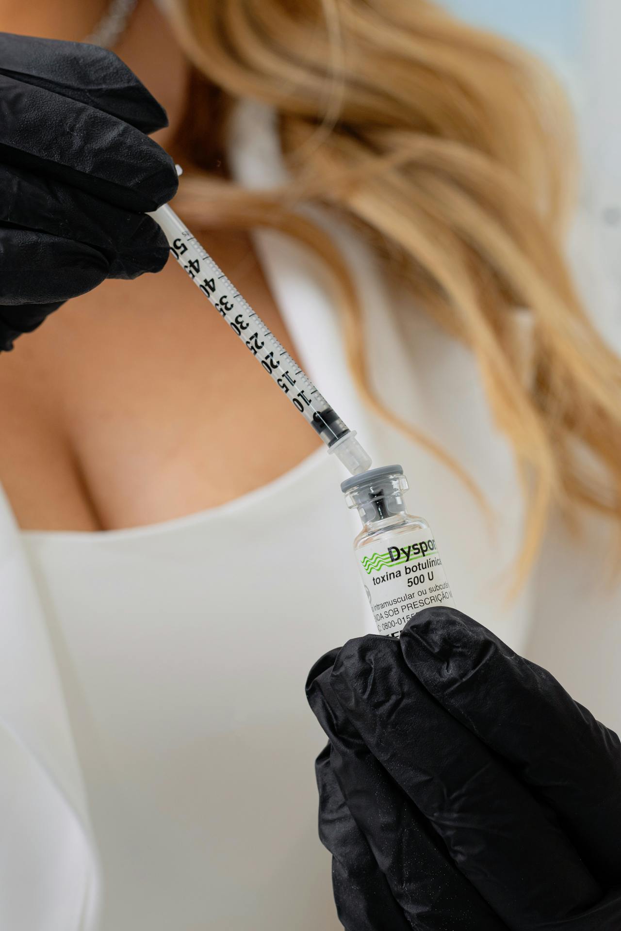
Botox temporarily relaxes the muscles that cause these movements. If the muscle isn’t contracting as forcefully, the skin isn’t folding as deeply or as often.
The idea behind preventive Botox is simple: if you reduce the repetitive folding before the lines become etched in, you may be able to delay or minimize the formation of deep static wrinkles—the kind that show up even when your face is completely at rest.
What the Research Shows: Is Botox Really preventive?
A 2022 evidence-based review published in the Journal of Cosmetic Dermatology looked at both clinical studies and real-world practice patterns for Botox in younger adults. The researchers reviewed high-quality randomized controlled trials and surveyed 141 aesthetic practitioners across multiple countries.
Here’s what they found: wrinkle prevention was the number one reason younger patients sought Botox treatment. Not correction—prevention. And the clinical data backed up the approach.
The study’s conclusion was clear: an individualized approach with lower doses for younger adults is recommended, and the clinical evidence supports Botox’s efficacy for both treatment and prevention in this age group. The researchers noted that younger patients who started treatment reported feeling “more attractive” and “more confident” compared to older patients who were trying to correct established lines.
What Dermatologists Actually See
There aren’t many 20-year controlled studies comparing identical twins where one got Botox and one didn’t. (Though actually, there is at least one famous case study of twins, and the difference is pretty striking.) What we do have is decades of clinical observation.
Dermatologists who have been practicing for 20+ years can look at their long-term patients and see patterns. Those who started treatments earlier tend to have less severe static lines later. The muscles themselves can actually weaken somewhat over years of treatment, meaning some patients need less product over time, not more.
Does this mean you have to start Botox at 25 or you’ve missed your window? Absolutely not. People see great results starting treatment at any age. But the mechanism behind prevention makes physiological sense, and the clinical observations support it.
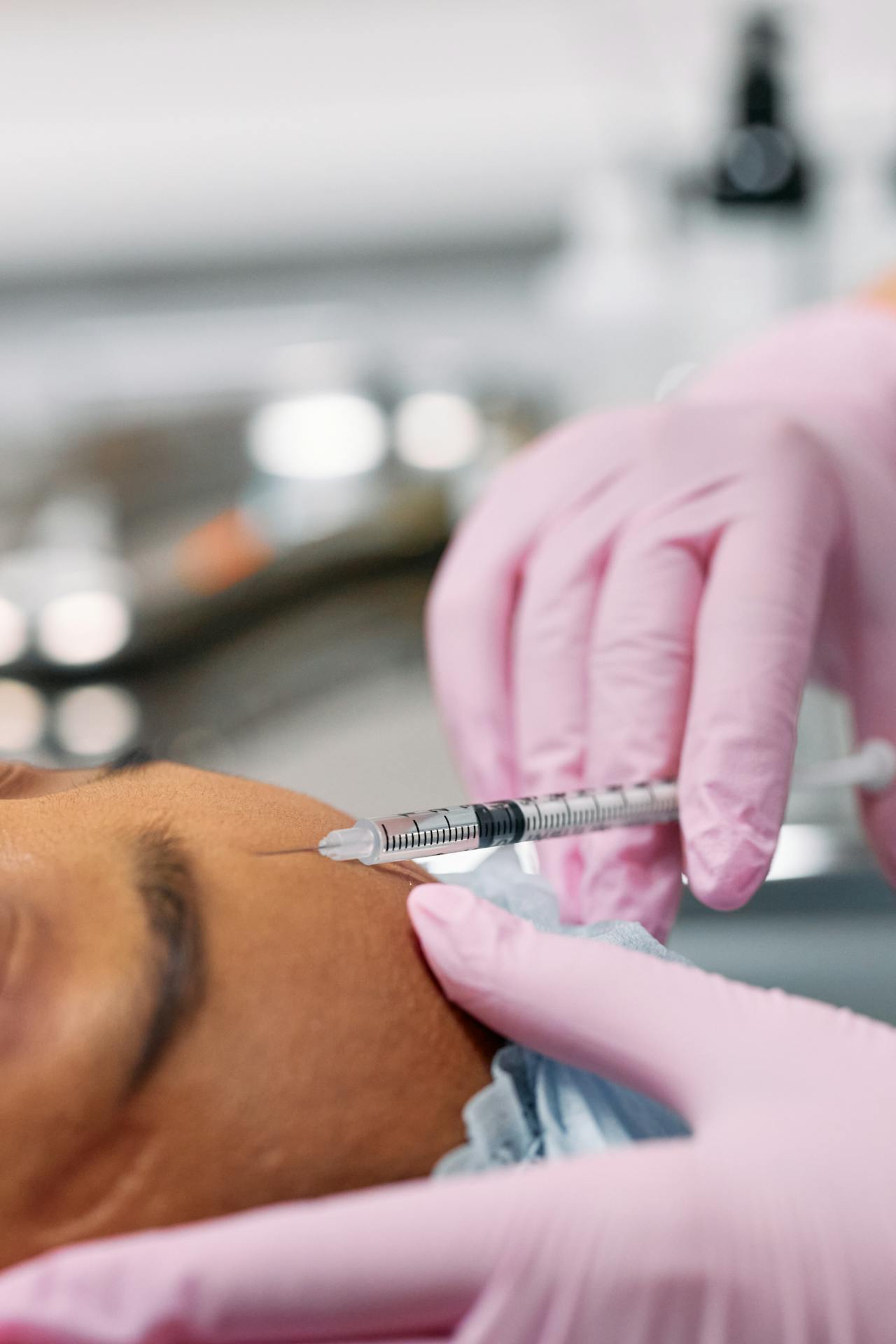
What preventive Botox Won’t Do
Here’s where the skeptics have some valid points, because preventive Botox does get oversold sometimes.
It won’t prevent wrinkles caused by sun damage. Those are about collagen breakdown from UV exposure, and Botox doesn’t address that. (Sunscreen does. Wear it.)
It won’t prevent volume loss—the gradual loss of fat and bone density in the face that happens as we age. Botox doesn’t add volume.
It won’t prevent skin laxity or sagging. That’s about skin structure, gravity, and genetics.
What Botox specifically addresses are dynamic wrinkles—the ones caused by muscle movement—and preventing those from becoming static wrinkles. That’s its thing. It’s one piece of a larger picture, and anyone telling you it’s a complete anti-aging solution isn’t being totally honest.
The Cost Question
Let’s talk about this because it’s usually what people are actually worried about underneath everything else.
Treatments typically run somewhere between $300-500 per session depending on what areas you’re treating, and results last around 3-4 months. That can add up.
Is it “a waste of money”? That really depends on what matters to you. People spend money on gym memberships, hair appointments, skincare routines, and plenty of other things that aren’t strictly necessary but make them feel good. Whether Botox fits into that category is pretty personal.
What’s worth knowing is that preventive doses are often smaller than corrective doses. Someone starting in their late 20s might need way less product than someone trying to soften lines that have been deepening for decades. So sometimes starting earlier actually means spending less per visit.
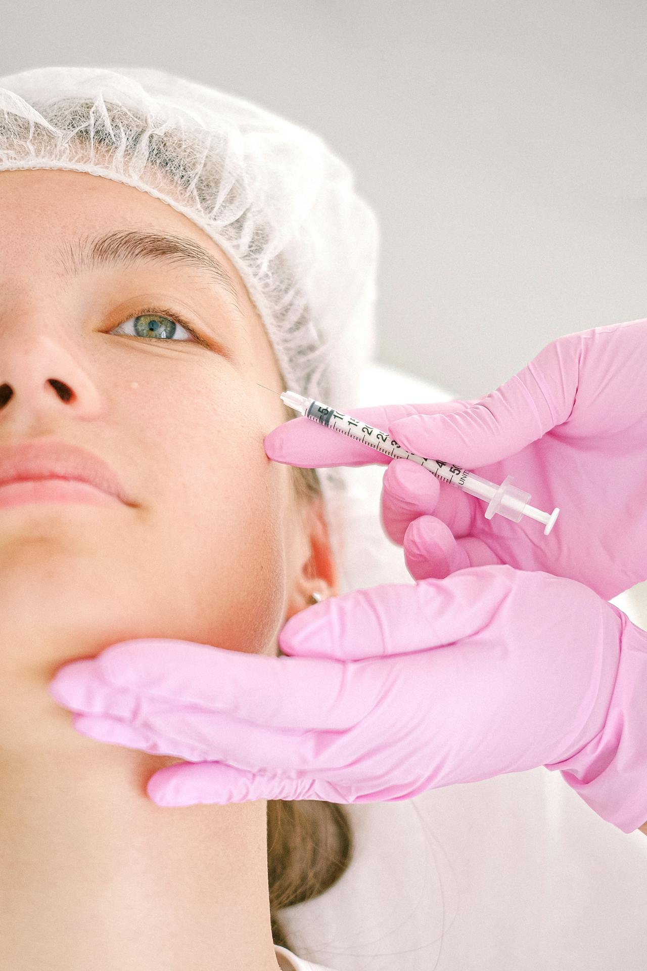
The Bottom Line
Preventive Botox isn’t a scam or some marketing scheme invented to get younger people into the chair. The mechanism is real and well-understood. Reducing repetitive muscle movements reduces repetitive skin folding, which can slow the formation of permanent creases. Clinical experience over decades backs this up.
It’s also not magic. Sun protection, genetics, overall health, skincare—all of that matters too. Botox is one tool, not the whole toolbox.
If you’ve been on the fence about it, or you’ve had someone in your life giving you a hard time about even considering it, hopefully this gives you something more concrete to work with. The best next step is always a conversation with a board-certified dermatologist who can look at your skin, your goals, and give you actual personalized advice instead of a generic sales pitch or a generic dismissal.
You deserve to make this decision with real information, not just opinions.
Aesthetic Dermatology Associates has offices in Media and Paoli, PA. We’re happy to chat through whether preventive or corrective Botox makes sense for you—no pressure, just answers.
Can I Workout After a Skin Biopsy? Managing Activity While Waiting for Results

Getting a skin biopsy can bring up two main concerns: how to care for the biopsy site and the anxiety that comes with waiting for results. If you’re wondering whether you can continue your normal routine—including exercise—and how to manage the days between your procedure and your phone call from the dermatologist, this guide covers what you need to know.
Returning to Exercise After a Skin Biopsy
Can I workout after a skin biopsy? For most patients, the answer is yes—with some sensible precautions.
Light to moderate exercise is usually fine the same day as your biopsy. Walking, stretching, and everyday activities won’t interfere with healing. However, there are a few things to consider:
For the first 48 hours: Keep the biopsy site dry and protected. This means avoiding swimming pools, hot tubs, and activities that cause heavy sweating. If your biopsy is on your leg or arm, high-impact exercises that strain or stretch the area should be postponed for a few days.
After 48 hours: Once you begin changing the bandage daily, you can gradually return to your normal workout routine. Just make sure to cleanse the site and apply fresh petroleum jelly and a clean bandage after exercising.
Listen to your body: If an activity causes pain, pulling, or bleeding at the biopsy site, scale back. Healing takes priority over maintaining your usual fitness schedule.
If you had a punch biopsy with stitches, ask your dermatologist about specific activity restrictions. Stitches may need extra care, and certain movements could delay healing or affect the final appearance of the scar.
How Long Should You Keep a Skin Biopsy Covered?
The initial bandage should stay in place for at least 48 hours. Keeping the site covered and dry during this period protects the wound and allows the earliest stage of healing to begin undisturbed.
After two days, you’ll start a simple daily routine: gently remove the old bandage, clean the area with mild soap and water, apply a thin layer of petroleum jelly, and cover it with a fresh bandage. Continue this for about one week, or until the site has closed and formed a protective layer.
Moist wound care—keeping the biopsy site slightly moist rather than letting it dry out—promotes faster healing and reduces the chance of noticeable scarring.
Skin Biopsy Healing Stages
Understanding the healing process can help set realistic expectations and reduce worry.
Days 1-2: The site may feel tender and show minor redness or swelling. A small amount of bleeding or oozing is normal. The bandage protects the area while initial clotting and tissue repair begin.
Days 3-7: A scab or thin crust usually forms. The surrounding skin may still appear slightly pink. This is the active healing phase, where new skin cells are regenerating beneath the surface.
Weeks 2-3: The scab naturally falls off on its own. The new skin underneath may look pink or lighter than the surrounding area. Avoid picking at the scab—letting it come off naturally minimizes scarring.
Weeks 4 and beyond: The color gradually evens out. Some biopsies leave a small, flat scar that continues to fade over several months. Protecting the area from sun exposure during this time helps prevent darkening or discoloration.
Why Does It Take So Long for Skin Biopsy Results?
One of the most common frustrations patients express is the waiting period. Results typically take 7 to 10 business days, and for many people, this feels like a long time.
Here’s what happens behind the scenes: After your dermatologist removes the tissue sample, it’s sent to a specialized pathology lab. A dermatopathologist—a physician trained to diagnose skin diseases under a microscope—examines thin slices of the tissue that have been processed and stained. This careful analysis takes time because accuracy is essential.
Some cases require additional steps, such as deeper staining techniques or a second pathologist’s review, which can extend the timeline by a few days. Weekends and holidays also affect turnaround time.
While waiting can be difficult, the thoroughness of this process ensures your dermatologist has the most accurate information to guide your care.
Nervous while waiting for biopsy results?

It’s completely normal to feel anxious while waiting for your results. Many patients describe checking their phone repeatedly, replaying the appointment in their mind, or worrying about what the biopsy might reveal.
A few things to keep in mind during this time:
Most biopsies come back benign. The majority of suspicious moles and lesions turn out to be non-cancerous. Common benign findings include seborrheic keratoses (harmless age-related growths), benign nevi (ordinary moles), and actinic keratoses (precancerous spots that are easily treatable).
Early detection is good news. If the biopsy does identify skin cancer—whether basal cell carcinoma, squamous cell carcinoma, or melanoma—finding it early means treatment is typically straightforward and highly successful.
You’ve already taken the most important step. By getting the biopsy done, you’re being proactive about your health. Waiting for results is part of the process, but you’ve already done the hardest part by seeking evaluation.
Avoid online searching. Every biopsy is unique. Reading about other people’s experiences or trying to self-diagnose based on internet descriptions usually increases anxiety rather than providing clarity.
If the waiting becomes overwhelming, reach out to your dermatologist’s office. A brief conversation with a staff member can often provide reassurance and remind you that help is available if needed.
What Happens Next
Once your results are ready, your dermatologist will contact you using the communication method you selected—phone call, secure message, or follow-up appointment. The reports will explain whether the lesion was benign, precancerous, or malignant, and outline any recommended next steps. For benign results, the biopsy report may only need to be reviewed at your next scheduled routine appointment.
If treatment is needed, options vary depending on the diagnosis. Non-cancerous findings may require no further action. Precancerous lesions might be treated with cryotherapy or topical medication. Skin cancers like basal cell carcinoma and squamous cell carcinoma are often removed with a simple excision. Melanoma may require more extensive evaluation and treatment, but early-stage melanoma has excellent cure rates.
When to Call Your Dermatologist
While complications after a skin biopsy are rare, contact your dermatologist if you notice:
- Bleeding that doesn’t stop after applying firm pressure for 10-15 minutes
- Increasing pain, redness, or swelling around the biopsy site
- Pus, unusual drainage, or a foul odor
- Fever or red streaks extending from the wound
These signs are uncommon, but it’s always better to check in early if something doesn’t seem right.
Moving Forward
A skin biopsy is a brief procedure with a clear purpose: to give your dermatologist the information needed to protect your health. The days that follow—caring for the site, adjusting your routine, and waiting for results—are manageable with the right information and a little patience.
Stay active within your comfort level, follow the aftercare instructions you received, and remember that most people who undergo skin biopsies receive reassuring results. If concerns arise, your dermatology team is available to help.
Other Articles You’d Want To Read
Mole Removal & Treatments: What You Need to Know
What to Do Right After a Skin Cancer Biopsy: First 48 Hours
Mohs Surgery or SRT? What Your Dermatologist Considers When Recommending Skin Cancer Treatment
Plaque Psoriasis: What Those Red Patches Mean (And Why There’s No Reason to Panic)
By Dr. Leib, Board-Certified Dermatologist – Aesthetic Dermatology Associates, serving Paoli & Media, PA
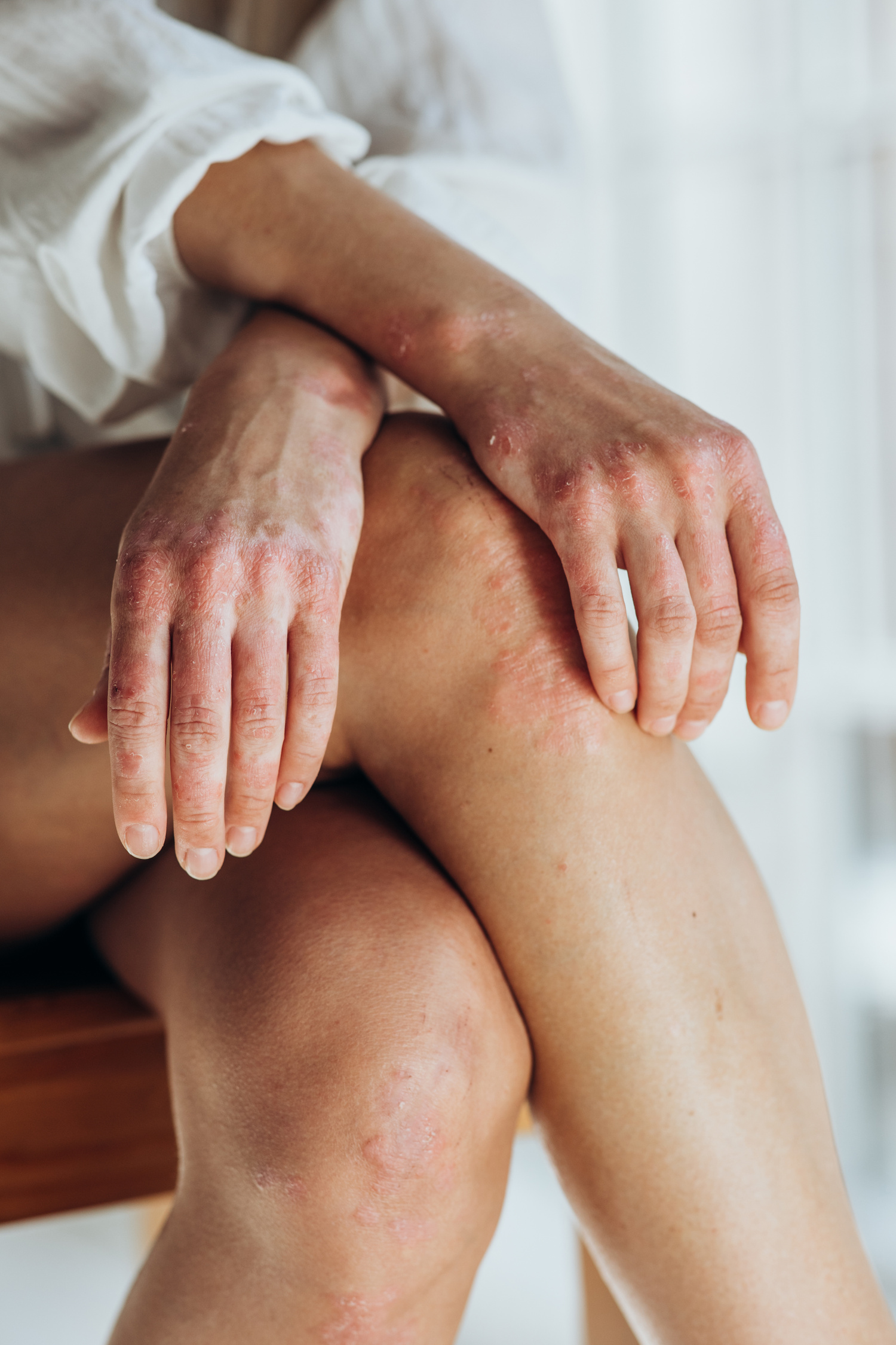
You noticed the first patch a few weeks ago. Maybe on your elbow, or your knee. Pink-red, slightly raised, with silvery-white scales on top. Your family doctor mentioned psoriasis, or maybe a friend recognized it from their own experience.
If you’re reading this because you suspect you have plaque psoriasis, you’re probably feeling anxious, confused, and maybe a little overwhelmed by everything you’re finding online. The good news is this: plaque psoriasis, while chronic, is manageable. Understanding what’s happening to your skin is the first step toward getting the right treatment and feeling more like yourself again.
Let’s start with the medical facts – explained in plain language.
What Is Plaque Psoriasis, Exactly?
Plaque psoriasis is a chronic inflammatory condition of the skin. Translation: it’s a long-term condition where your immune system causes inflammation.
Those raised patches you’re seeing? Dermatologists call them “plaques.” They’re typically pink-red in color with whitish or silvery scales on top. They can appear almost anywhere on your body, but most commonly show up on:
- Knees and elbows (the most common spots)
- Scalp
- Lower back
- Underarms and groin
- Buttocks
- Nails (causing pitting, discoloration, or thickening)
Dr. Leib explains:
“When patients first come in with suspected psoriasis, one of the first things I tell them is: this is not something you caused. It’s not from poor hygiene, something you ate, or anything you did wrong. Plaque psoriasis is an immune-mediated condition – meaning your immune system is the driving force behind what you’re seeing on your skin.”
What Causes Plaque Psoriasis? The Real Answer
This is probably the question weighing most heavily on your mind right now. And here’s the honest answer: like so many medical conditions, the cause of psoriasis is complex. We generally cannot blame one particular thing.
The genetic component:
If you have plaque psoriasis, you have a genetic predisposition. This means you inherited genes that make your immune system more likely to behave this way. You might have family members with psoriasis, or you might be the first person in your family to develop it – both scenarios are common.
Having the genetic tendency doesn’t mean you’ll definitely develop psoriasis. But it does mean that under the right circumstances, your immune system can shift into this overactive state.
Environmental triggers:
Even with genetic susceptibility, something usually acts as a trigger – an environmental factor that essentially “activates” the condition. Common triggers include:
- Stress: Emotional or physical stress is one of the most significant triggers
- Trauma to the skin: Cuts, scrapes, sunburns, or even tattoos can trigger plaques to form at the injury site
- Infections: Strep throat and other infections can trigger psoriasis, especially in younger people
- New medications: Certain drugs, including lithium, beta-blockers, and some blood pressure medications, can trigger or worsen psoriasis
- Weather changes: Cold, dry weather often triggers flares
- Lifestyle factors: Smoking and heavy alcohol use can contribute to development and severity
Dr. Leib emphasizes:
“Many patients come in trying to figure out exactly what caused their psoriasis. While identifying triggers can be helpful for managing the condition, it’s important not to blame yourself. Sometimes we can identify a clear trigger, and sometimes we can’t. Either way, what matters most is moving forward with effective treatment.”
How Common Is Plaque Psoriasis?
You’re far from alone. About 2% of the world’s population is affected by psoriasis, though the frequency is thought to be higher in the United States. That’s millions of people living with this condition.
Psoriasis can occur at any age – from infancy to late adulthood. However, there are two peak times when people are most likely to develop it:
- Ages 20-30 (the most common time for first onset)
- Ages 50-60 (the second peak period)
If you’re being diagnosed in your twenties or thirties, you might feel frustrated that this is happening when you’re supposed to be at your healthiest. But this timing is actually completely typical for plaque psoriasis.
What Does Plaque Psoriasis Feel Like? Recognizing the Symptoms
Unlike eczema (another common inflammatory skin condition), plaque psoriasis tends to be only mildly itchy. Some people experience virtually no itching at all, while others find it bothersome but not severe.
The most common symptoms include:
- Dry, cracked skin
- Stinging or burning sensations, especially if the skin cracks or fissures
- Joint pain or stiffness (if psoriatic arthritis is developing)
A word about cracks and fissures:
Occasionally, psoriasis plaques can cause cracks or fissures in the skin. These can sting, burn, or become painful – especially in areas that bend or flex frequently. These cracks also create an opening for bacteria, which can lead to infections.
If you notice increased warmth, swelling, oozing, or fever alongside your psoriasis, contact your dermatologist right away – these could be signs of infection requiring prompt treatment.
The Psoriatic Arthritis Connection
Here’s something important to know: plaque psoriasis can be associated with psoriatic arthritis – a condition that causes painful inflammation in the joints.
Not everyone with plaque psoriasis will develop psoriatic arthritis, but the two conditions are connected. If you’re experiencing joint pain, stiffness (especially in the morning), or swelling alongside your skin symptoms, mention this to your dermatologist. Catching psoriatic arthritis early and treating it promptly can help prevent permanent joint damage.
Will Plaque Psoriasis Go Away on Its Own?
This is one of the most common questions newly diagnosed patients ask, and it’s important to be honest about the answer.
Even though symptoms aren’t necessarily severe, plaque psoriasis is very stubborn and usually does not go away without treatment. This is one of the key differences between psoriasis and other skin conditions.
Dr. Leib notes:
“I understand that hearing ‘chronic condition’ can feel discouraging. But here’s what that really means: psoriasis is something we manage, not something we cure. And the good news is that we have many, many effective treatment options available today. Although there is no cure for this condition, thankfully there are many treatments that can help minimize the appearance of the plaques and alleviate symptoms. Many of my patients achieve clear or nearly clear skin with the right treatment plan.”
What Happens Next: Your Path Forward
If you’re reading this because you suspect you have plaque psoriasis, the next step is to see a board-certified dermatologist who can:
- Confirm the diagnosis: While plaque psoriasis has characteristic features, other conditions can sometimes look similar. A dermatologist can examine your skin and, if needed, perform a simple biopsy to confirm the diagnosis.
- Assess the severity: Not all psoriasis is the same. Your dermatologist will evaluate how much of your body is affected and how the condition is impacting your quality of life.
- Create a personalized treatment plan: From prescription topical treatments to light therapy to advanced biologic medications, there are numerous options. Your treatment plan will be tailored to your specific situation, preferences, and lifestyle.
- Provide ongoing support: Psoriasis management is a partnership. Your dermatologist will work with you over time to adjust treatments as needed and help you identify and avoid your personal triggers.
You’re Not Alone in This
Finding out you have a chronic skin condition can feel isolating. You might worry about how it looks, whether it will get worse, or how it will affect your daily life.
But here’s what you need to know: plaque psoriasis is highly treatable. With the right dermatology partner and a treatment plan designed for you, most patients see significant improvement. Many achieve clear or nearly clear skin and regain the confidence that psoriasis initially took away.
This condition doesn’t have to define you or limit your life. Treatment works. Relief is possible. And you don’t have to figure this out alone.
About Dr. Leib
Dr. Leib is a board-certified dermatologist with extensive experience treating plaque psoriasis and other complex skin conditions in southeastern Pennsylvania. She served as chief resident during her dermatology training at Virginia Commonwealth University and has contributed to the field through peer-reviewed publications and presentations at regional dermatology conferences.
A Downingtown native and Penn State College of Medicine graduate, Dr. Leib is an active member of leading dermatology specialty societies. She is dedicated to patient-centered care, staying current with the latest treatment advances, and providing compassionate, expert guidance to every patient navigating their psoriasis journey.
Related links:
See Dr. Leib’s provider’s page on our website https://adaderm.com/providers/amy-leib-md/
Review 5-star rating given by patients https://share.google/wJjqWA8m5OvPfw6Hx
Schedule Your Plaque Psoriasis Consultation in Paoli or Media, PA
Understanding what’s happening to your skin is the first step. Getting expert care is the next.
At Aesthetic Dermatology Associates, Dr. Leib and our experienced team offer:
- Accurate diagnosis: Comprehensive evaluation to confirm plaque psoriasis and rule out other conditions
- Personalized treatment plans: Tailored to your lifestyle, severity, and treatment goals
- Full range of treatment options: From prescription topicals to advanced biologic therapies
- Compassionate, expert care: A dermatology partner who understands your concerns and guides you every step of the way
Conveniently serving patients in Paoli, PA and Media, PA
Don’t let uncertainty keep you from getting the care you need. Whether you’re newly diagnosed or seeking better management, we’re here to help you achieve clear, comfortable skin – and peace of mind.
Rosacea on Your Face: What That Redness Means (And Why There’s No Reason to Panic)

By Nancy Conley, PA-C, Diplomate of the Society of Dermatology Physician Assistants – Aesthetic Dermatology Associates, serving Paoli & Media, PA
Your face has been turning red more than usual. At first, you thought it was just from the heat, or maybe that glass of wine at dinner. But now the redness lingers. Your cheeks seem perpetually flushed, and you’ve noticed small bumps that almost look like acne – except your go-to acne cream isn’t helping at all.
Maybe your doctor mentioned the word “rosacea” during your last visit. Or perhaps a friend who has it recognized the telltale signs. Now you’re online at night, scrolling through images and articles, asking yourself:
Is this really what’s happening to my face? What caused this? And can I make it go away?
If you’re here because you think you might have rosacea, you’re likely feeling self-conscious, frustrated, and worried about what this means for your skin going forward. Here’s what you need to hear right now: rosacea is one of the most common facial skin conditions, and it responds incredibly well to proper treatment. You’re not stuck with persistent redness and sensitivity. Understanding what’s causing your symptoms is the first step toward skin that looks and feels healthy again.
Let’s talk about what’s really happening – in terms that actually make sense.
The Truth About Rosacea: It’s More Than Just Blushing

Rosacea is a chronic inflammatory skin condition that primarily affects the face. In practical terms, this means your facial blood vessels and oil glands react more intensely than they should to certain triggers, causing visible redness, flushing, and sometimes acne-like breakouts.
Here’s what makes rosacea different from just having “sensitive skin” or occasional flushing:
The symptoms persist. That rosy glow or facial redness doesn’t fully fade the way a normal flush would. For many people, there’s a baseline redness that’s always there, punctuated by episodes where it gets much worse.
The symptoms come and go in waves. You might have weeks where your skin looks relatively calm, followed by a flare-up where everything gets worse – more redness, more bumps, more discomfort. These flare-ups are usually triggered by specific factors (more on that in a moment).
You might be experiencing:
- Redness that won’t fade, especially across your nose, cheeks, chin, and forehead
- A face that flushes easily and intensely
- Tiny red or purple blood vessels visible just under your skin
- Bumps and small pimples that appear without warning
- Skin that stings, burns, or feels uncomfortably tight
- Rough, dry patches even when you’re moisturizing
- A sensation of heat in your face
- Eyes that feel irritated, dry, or bloodshot
Nancy Conley explains:
I see patients every day who are self-conscious about their red, acne prone skin. When I tell them it’s rosacea and I can treat it, there is a moment of relief. Then we jump into the nitty gritty – what it is and how I can help.
The Trigger Puzzle: Why Your Face Flares Up
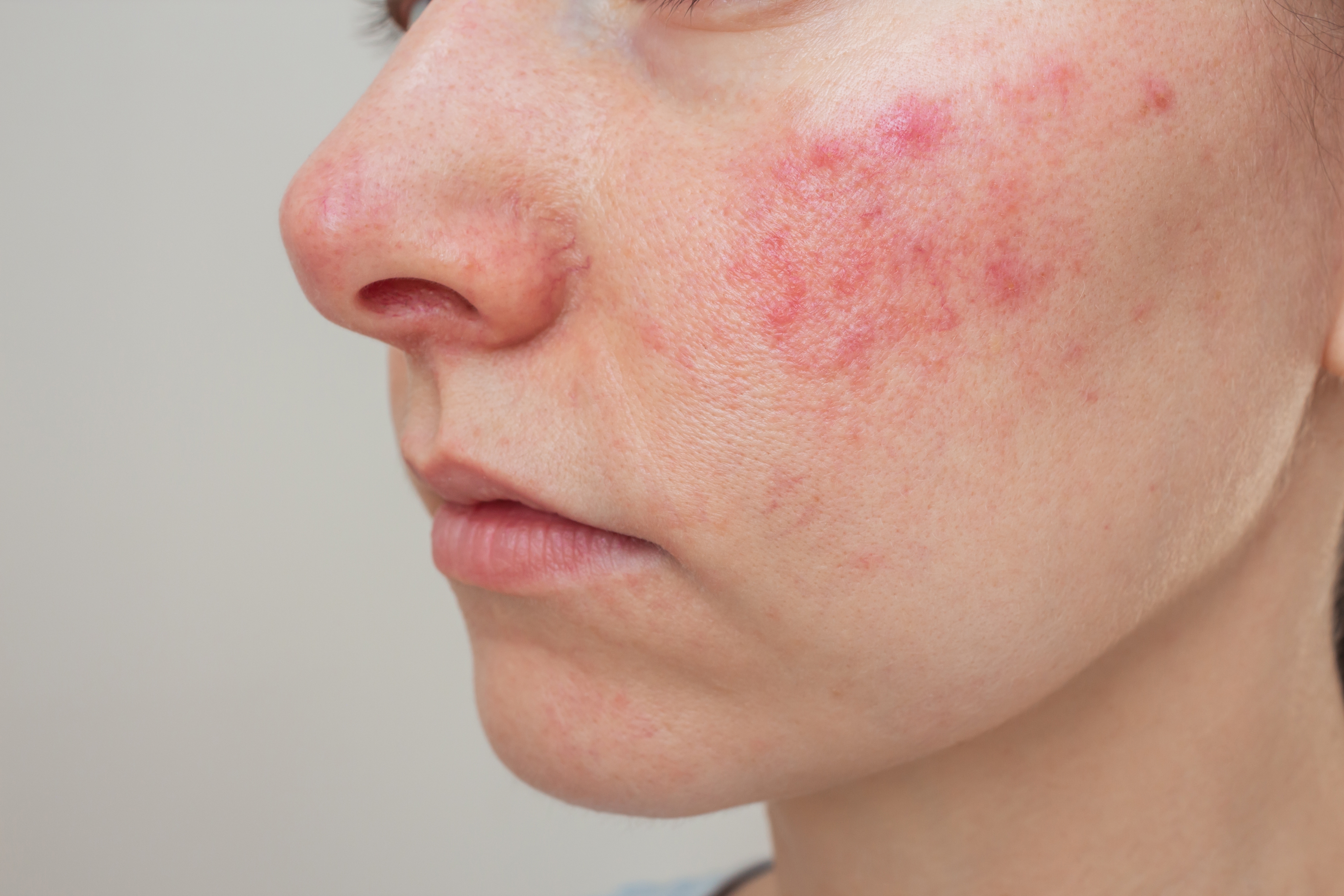
Even though we can’t always prevent rosacea from developing, we can absolutely identify and manage the things that make it worse. These are called triggers, and they’re different for everyone.
The most common culprits include:
Sun exposure: This is the number one trigger for most people with rosacea. UV rays cause blood vessels to dilate and inflammation to increase. Even brief sun exposure can cause a flare that lasts for days.
Alcohol consumption: Wine (especially red wine), beer, and spirits all commonly trigger facial flushing and redness in people with rosacea.
Spicy foods: Anything with heat – hot peppers, curry, hot sauce – can cause your face to flush and stay red for hours.
Temperature extremes: Very hot or very cold environments, or moving quickly between them (like going from air conditioning into summer heat), can trigger symptoms.
Hot beverages: That morning coffee or afternoon tea might be contributing to your facial redness, especially if you drink it while it’s very hot.
Stress and strong emotions: Emotional stress, anxiety, anger, or embarrassment can all cause flushing and worsen rosacea.
Harsh skincare products: Products with fragrances, alcohol, exfoliating acids, or rough physical scrubs can irritate rosacea-prone skin and trigger flares.
Intense exercise: While exercise is healthy, the flushing and heat it generates can worsen rosacea for some people.
Living With Rosacea: What It Actually Feels Like Day-to-Day
Beyond the visible redness and bumps, rosacea affects how your skin feels and how you feel about your skin.
Physically, many people describe:
- A burning or stinging sensation, especially when applying products
- Tightness, like your skin is too small for your face
- Constant awareness of warmth in your cheeks or nose
- Discomfort that gets worse throughout the day
- Sensitivity to products you used to tolerate just fine
Emotionally, rosacea can take a toll:
- Self-consciousness about your appearance
- Frustration when flare-ups happen unpredictably
- Anxiety about social situations where you might flush
- Feeling like you have to explain your “red face” to people
- Avoiding activities (like hot yoga or spicy dinners) that you used to enjoy
These feelings are completely valid. Your skin is on your face – the most visible part of your body. Of course it affects how you feel. This is exactly why getting proper treatment isn’t vanity; it’s taking care of your well-being.
The Bottom Line: Will It Get Better Without Treatment?
Here’s what you need to know: rosacea rarely improves on its own. In most cases, without treatment, it actually gets progressively worse over time.
What might start as occasional flushing can evolve into permanent redness. Small bumps can become more frequent and widespread. In some cases, the skin can thicken, particularly around the nose.
Nancy Conley is direct about this:
I never want to scare patients, but I want to them to understand control is attainable. While there is no cure for this condition, thankfully many treatments are available to give you your best skin. Whether you’ve only had symptoms for a few months, or even symptoms for many years, we can achieve significant improvement and prevent progression!
Building Your Rosacea-Friendly Skincare Routine
Before prescription treatments, let’s talk about the foundation: how you care for your skin every single day. For rosacea, gentle skincare isn’t optional – it’s essential.
The golden rule: sensitive skin care is key.
Your skin barrier is compromised when you have rosacea. It’s more reactive, more sensitive, and less able to tolerate products and ingredients that other people use without issue. This means you need to completely rethink your skincare approach.
Products to avoid completely:
- Anything fragranced, even if it says “natural fragrance”
- Exfoliants of any kind – no scrubs, no acids, no chemical peels
- Alcohol-based toners or astringents
- Witch hazel
- Menthol, peppermint, eucalyptus, or other “cooling” or “tingling” ingredients
- Rough washcloths, loofahs, or cleansing brushes
- Hot water on your face
What your face wash should be:
Choose a gentle facial cleanser specifically formulated for sensitive skin. Look for something creamy, non-foaming, and fragrance-free. Alternatively, Nancy Conley recommends a medicated sulfur cleanser, which can help calm inflammation while still cleansing effectively.
Wash with lukewarm (not hot) water, using only your fingertips. Pat dry gently with a soft towel – don’t rub.
When your skin is flaring:
A hydrating face mask can provide immediate relief during a flare-up. Nancy specifically recommends Skinceuticals Phyto Corrective Mask for its ability to instantly calm irritated, inflamed skin. Keep it in your skincare arsenal for those days when your face feels particularly angry.
Sun protection – non-negotiable:
Every single day, rain or shine, winter or summer, you need sunscreen. Choose a mineral-based formula (zinc oxide or titanium dioxide) with at least SPF 30. Chemical sunscreens can sometimes irritate rosacea-prone skin, so mineral formulas are generally safer.
Moisturizer matters:
Even if your skin feels oily or you have acne-like bumps, you need a good moisturizer. Choose something designed for sensitive skin, fragrance-free, and with soothing ingredients like ceramides or niacinamide.
Medical Treatments That Make a Real Difference
The right skincare routine sets the foundation, but for most people with rosacea, medical treatment is what really transforms the skin.
Topical prescription creams:
Multiple prescription medications can reduce redness, calm inflammation, and minimize bumps and visible blood vessels. These are typically applied once or twice daily and work by targeting the specific inflammatory processes happening in rosacea-affected skin.
Oral antibiotics:
For moderate to severe rosacea, especially when you have significant bumps and pustules, oral antibiotics can provide dramatic improvement. These work by reducing inflammation throughout your body, not by fighting infection. They’re typically used for a limited time to get symptoms under control.
Laser and light treatments:
Advanced laser therapies can specifically target the visible blood vessels and persistent background redness that topical treatments sometimes can’t fully address. These treatments provide long-lasting improvement and can significantly reduce the overall redness of your complexion.
Why one-size-fits-all doesn’t work:
Your rosacea is unique to you. The severity, the specific symptoms, your skin type, your lifestyle, your budget, your treatment preferences – all of these factors matter when creating a treatment plan.
Nancy Conley explains her approach:
When I sit down with a rosacea patient, I want to understand the whole picture. What bothers you most – the redness, the bumps, the sensitivity? What have you already tried? What’s your daily routine like? Are you willing to take an oral medication, or would you prefer to stick with topical treatments? Do you want to consider laser therapy? It’s always best to consult with your dermatology provider to discuss treatment options that would be best for your particular skin. This is a collaborative process, and my job is to help you find the approach that works for your life and your goals.
Taking the Next Step: Getting Professional Help
If you’ve read this far and you’re recognizing yourself in these descriptions, it’s time to see a dermatology provider who can give you a definitive diagnosis and start you on the path to clearer skin.
Here’s what to expect when you come in for a consultation:
Comprehensive skin evaluation: Your provider will examine your face closely, ask about your symptoms and their timeline, and discuss your skincare routine and lifestyle factors. In most cases, rosacea can be diagnosed based on appearance and history alone, without any testing needed.
Customized treatment strategy: Based on your specific symptoms, skin type, and preferences, your provider will recommend a treatment plan. This might include skincare changes, prescription medications, laser treatments, or a combination of approaches.
Ongoing partnership: Rosacea is a chronic condition, which means your treatment might need adjustments over time. Your dermatology provider will follow your progress, modify your treatment as needed, and help you navigate challenges as they come up.
You don’t have to live with constant redness and sensitivity. You don’t have to avoid activities you love or feel self-conscious about your appearance. Treatment works. Relief is real. And you absolutely don’t have to figure this out alone.

About Nancy Conley, PA-C
Nancy Conley, is a board-certified Physician Assistant specializing in medical and cosmetic dermatology, with particular expertise in treating rosacea and sensitive skin conditions. As a Diplomate of the Society of Dermatology Physician Assistants (SDPA) – a distinction earned by only a select group of PAs nationwide – Nancy has completed advanced dermatology-specific training that enables her to provide expert care for even the most challenging rosacea cases.
A Philadelphia-area native and proud member of the Aesthetic Dermatology Associates team, Nancy is known for her patient-centered approach to rosacea treatment. She takes time to understand each patient’s unique triggers, lifestyle factors, and treatment goals, creating customized care plans that deliver real results. Licensed in Pennsylvania, Nancy combines clinical excellence with genuine compassion, helping patients achieve clear, calm, confident skin.
Related link:
See Nancy’s provider’s page on our website.
Get Expert Rosacea Care in Paoli or Media, PA
Understanding what’s happening to your face is empowering. Getting the right treatment is life-changing.
At Aesthetic Dermatology Associates, Nancy Conley and our experienced team provide:
- Accurate rosacea diagnosis: Thorough evaluation to confirm rosacea and identify your specific subtype
- Customized treatment plans: Designed around your unique skin, lifestyle, and goals
- Complete range of solutions: From gentle skincare guidance to prescription medications to advanced laser therapies
- Expert, compassionate care: A partner who listens to your concerns and supports you through every step of your treatment journey
Conveniently located to serve patients throughout Paoli, PA and Media, PA
Don’t let facial redness and sensitivity hold you back from feeling confident in your skin. Whether you’re just noticing symptoms or you’ve been struggling with rosacea for years, we’re here to help you achieve the clear, calm, comfortable skin you deserve.
Visit Our Locations
Media, PA
Media, PA 19063
Phone hours may differ.
| Mon: | 7:30 am - 5:30 pm |
| Tue: | 7:30 am - 4:30 pm |
| Wed: | 7:30 am - 4:30 pm |
| Thu: | 7:30 am - 4:30 pm |
| Fri: | 7:30 am - 4:30 pm |
Paoli, PA
Paoli, PA 19301
Phone hours may differ.
| Mon: | 7:30 AM - 4:30 PM |
| Tue: | 7:30 AM - 4:00 PM |
| Wed: | 7:30 AM - 3:45 PM |
| Thu: | 7:30 AM - 4:30 PM |
| Fri: | 7:30 AM - 3:30 PM |
Stay Informed with our Newsletter

Book Your Appointment Today!
Choose the option that works best for you:
Prefer to speak with someone? Call or text us:

Media, PA Office - Call or Text (610) 566-7300
Paoli, PA Office - Call or Text (610) 566-7300
Want to book online now? Select a provider below:
Note: Self-scheduling is currently available for Dr. Goggins & Dr. Waller. For all other providers, please call or text the office.